Bow Leg Correction

Home / Treatments / Bow Leg Correction

Bow Leg Correction

Home/Treatments/Bow Leg Correction

What are bow legs?

Bowlegs, medically termed genu varum, is a structural condition in which the legs curve outward at the knees while the ankles and feet remain together. This creates a visible gap between the knees when a person stands upright. It’s often observed in infants and toddlers as part of normal development, but if it persists beyond early childhood, it may indicate an underlying pathological cause.

In its essence, bow legs represent a misalignment in the mechanical axis of the lower limbs. Instead of forming a straight line from the hips to the ankles, the legs bow outward, disrupting normal biomechanical function. This malalignment can exert abnormal stress on the knees, hips, and ankles, eventually leading to joint degeneration, gait abnormalities, and chronic discomfort.

Differences Between Physiological and Pathological Bowlegs

Physiological bowlegs are a common occurrence in infants and typically resolve by the age of 2–3 years as the child grows and their legs straighten. This is due to natural changes in bone growth and weight-bearing development. In contrast, pathological bow legs persist beyond this age or appear suddenly in older children or adults. These may result from various conditions, including nutritional deficiencies, bone diseases, or genetic disorders.

Key differentiators include:

  • Age of onset: Physiological typically appears from birth to 2 years; pathological can develop later.
  • Symmetry: Physiological cases are often symmetrical; pathological cases may show unilateral deformity.
  • Progression: Physiological bowing improves with age, while pathological bowing may worsen or remain unchanged.
  • Associated symptoms: Pain, stiffness, or limping indicate a pathological origin.

Recognising the type is crucial because while physiological bowing often requires observation only, pathological cases may need active intervention.

Age Groups Commonly Affected

Bow legs are most common in:

  • Infants and toddlers (0–3 years): Usually physiological and self-correcting.
  • Children aged 3–10 years: Pathological causes such as Blount’s disease or rickets may begin to manifest.
  • Adolescents and adults: Typically indicative of untreated childhood conditions, degenerative diseases like osteoarthritis, or trauma-induced deformities.

In adults, bow legs often present more significant challenges due to the mature skeletal structure and associated joint damage. Surgical correction becomes more relevant at this stage, particularly if mobility is compromised or pain is significant.

Causes of Bowlegs

Developmental Phase in Infants and Toddlers

During infancy, bowing of the legs is completely normal. As children begin to walk and bear weight, the mechanical stress on their legs encourages straightening. Most children outgrow this phase by 2–3 years old. This natural correction is due to bone remodelling during growth.

This physiological bowing doesn’t require treatment unless it persists beyond age 3 or worsens, which would then prompt further investigation.

Genetic and Hereditary Influence

Genetics can play a strong role in determining leg alignment. Children with a family history of bowed legs or skeletal abnormalities may be more likely to exhibit similar patterns. In such cases, the condition may persist beyond the expected correction age or reappear during adolescence.

Hereditary conditions such as achondroplasia or other skeletal dysplasias can also present with bowlegs as a primary symptom, often alongside short stature and other skeletal anomalies.

Nutritional Deficiencies (Vitamin D and Rickets)

One of the most well-known causes of pathological bowlegs is rickets, a disease resulting from severe vitamin D deficiency. Vitamin D is essential for calcium absorption and bone mineralisation. When deficient, bones become soft and prone to deformities under the mechanical stress of walking.

Signs of rickets-related bowlegs often include:

  • Delayed walking
  • Muscle weakness
  • Bone pain or tenderness
  • Widened wrists or ankles

Rickets can be prevented with adequate sun exposure, a balanced diet rich in vitamin D and calcium, and supplementation when necessary. In some cases, orthopedic intervention may still be required to correct deformities even after nutritional repletion.

Underlying Medical Conditions (Blount’s Disease, Osteoarthritis)

Blount’s disease is a growth disorder of the tibia (shin bone) that causes the lower leg to angle inward, worsening bowing. It is typically diagnosed in children or adolescents and may require bracing or surgery depending on severity.

In adults, osteoarthritis, particularly of the medial knee compartment, can lead to progressive bowing of the legs due to joint space narrowing, cartilage degradation, and bone remodelling. Abnormal alignment exacerbates joint damage, which in turn increases deformity, creating a vicious cycle.

Other conditions like skeletal dysplasias, metabolic bone diseases, and post-traumatic growth disturbances can also lead to genu varum.

Symptoms and Signs of Bow Legs

Outward Curvature and Posture Issues

The hallmark symptom of bow legs is the visual curvature of the legs, especially evident when standing with feet together and knees apart. In mild cases, the deformity may be subtle and asymptomatic. However, as the condition progresses, the curvature becomes more pronounced, affecting leg symmetry and body alignment.

This misalignment places abnormal stress on the inner (medial) aspect of the knees and can lead to compensatory postural issues. Over time, this can affect spinal alignment, pelvic tilt, and even shoulder posture.

Patients may also report feeling unbalanced or unstable while standing or walking, often describing a “waddling” gait pattern.

Functional Impairments (Pain, Gait Problems, Joint Stiffness)

As bow legs advance, functional issues begin to appear:

  • Pain: Commonly in the knees, hips, or ankles due to uneven weight distribution.
  • Fatigue: Especially after walking or standing for long periods.
  • Gait disturbance: Including a broad-based or waddling gait.
  • Stiffness: Especially during movement after rest.
  • Uneven shoe wear: Due to abnormal foot mechanics and pressure distribution.
  • Balance issues: Resulting from an altered center of gravity.

In children, these signs may manifest as delayed walking, reluctance to run or jump, or frequent falls. In adults, symptoms may be mistaken for generalized arthritis until a proper orthopedic assessment is conducted.

When to See an Orthopaedic Specialist - Dr Divya Ahuja

Persistence Beyond Early Childhood

If bowlegs persist beyond age 3 or worsen rather than improve, it’s essential to consult an orthopaedic specialist. Persistent bowing may indicate an underlying bone disorder or require corrective intervention to prevent long-term damage.

Recognising Signs in Adolescents and Adults

Adolescents and adults developing bow legs—or those noticing progression—should seek evaluation. Often, bowing at this stage reflects cumulative joint stress, trauma, or degenerative changes such as osteoarthritis.

Key signs to watch for:

  • Pain during activity
  • Increased curvature over time
  • Limitations in physical movement
  • Poor balance or stability
Mobility Issues or Family History

Patients experiencing mobility difficulties, joint locking, or weakness should not delay medical attention. Similarly, a family history of rickets, Blount’s disease, or skeletal deformities increases the risk and warrants closer monitoring of children during growth phases.

Effects of Untreated Childhood Conditions

Failure to treat pathological bow legs during childhood can result in a cascade of complications in adulthood, including:

  • Chronic joint pain
  • Degenerative arthritis
  • Limb length discrepancies
  • Abnormal gait and posture
  • Decreased physical performance

These consequences underline the importance of early detection and appropriate intervention, especially in high-risk children.

Diagnosis of Bow Legs

Clinical Examination and Medical History

Accurate diagnosis begins with a comprehensive clinical examination and detailed medical history. The orthopaedic specialist evaluates leg alignment while the patient is standing and walking. They observe the presence and degree of outward curvature, assess the symmetry of both legs, and look for any signs of joint instability or muscle weakness.

Key points in the patient’s history include:

  • Age of onset
  • Family history of skeletal conditions
  • Past nutritional deficiencies or growth abnormalities
  • Onset and pattern of symptoms (e.g., pain, stiffness, limping)

The physical examination helps determine if the condition is physiological (likely to resolve with growth) or pathological (requires further evaluation and treatment).

Imaging Tests and Measurement of Deformity

After physical evaluation, radiographic imaging (X-rays) is essential. It helps quantify the deformity and identify its location—whether in the femur (thigh bone), tibia (shin bone), or both.

  • Full-length standing X-rays: Show the mechanical axis of the leg, i.e., the line from the hip to the ankle that normally passes through the centre of the knee.
  • MRI or CT scans: Occasionally used to assess joint cartilage, ligament integrity, or underlying bone conditions.

Important diagnostic angles include:

  • Tibiofemoral angle: Measures the degree of bowing.
  • Mechanical axis deviation (MAD): Indicates whether the deformity is localised or affecting multiple segments.

These measurements help orthopaedic surgeons determine the exact site of deformity and plan appropriate treatment strategies.

Treatment Options for Bowlegs

Non-Surgical Treatments

In children with physiological bowing, no intervention may be needed. Instead, the orthopaedic specialist may suggest:

  • Observation: Routine follow-ups to monitor the natural correction of leg alignment over time.
  • Physical therapy: Strengthens leg muscles, improves joint mobility, and enhances gait. Custom exercises target the hip abductors, quadriceps, and core stabilisers.
  • Orthotic devices or braces: Used to gradually realign the legs. Young children, whose bones are still growing, benefit more from these exercises.
  • Nutritional therapy: Especially crucial in cases of rickets. Treatment includes:
    • Vitamin D supplementation (cholecalciferol or ergocalciferol)
    • Calcium and phosphate correction
    • Exposure to sunlight

These noninvasive methods can yield significant improvement in early-stage or mild cases. However, in adolescents and adults, where bone growth has completed, conservative treatments are usually ineffective for significant deformity.

Surgical Treatments

For moderate to severe deformities or when non-surgical options fail, surgical intervention is the definitive treatment.

Osteotomy:

  • Osteotomy is the most commonly performed surgical procedure for bowlegs.
  • The affected bone (femur or tibia) is cut and realigned to correct the mechanical axis.
  • May be accompanied by internal fixation using plates, rods, or screws.
  • This procedure is typically chosen for younger adults who have healthy bones.

External Fixation Devices:

  • Involves attaching a circular or rod-based frame around the leg.
  • This device enables the gradual correction of deformity through adjustable struts.
  • Beneficial in complex or multi-planar deformities.

Guided Growth Surgery:

  • Growing children can undergo this minimally invasive procedure.
  • A small metal plate is placed on one side of the growth plate to guide the bone to grow straighter.
  • If timed correctly, this procedure is reversible and highly effective.

Limb Lengthening:

  • This procedure may be necessary if there is a discrepancy in leg length.
  • An external fixator or an intramedullary lengthening nail is used to perform this procedure.

Total Knee Replacement (TKR):

  • Older adults with severe bow legs complicated by advanced osteoarthritis may consider Total Knee Replacement (TKR).
  • TKR realigns the joint surfaces, relieves pain, and restores function.

Each surgical plan is individualised based on the patient’s age, degree of deformity, symptoms, and overall health status.

Benefits of Bow Leg Correction

Structural and Functional Improvements

Correcting bowlegs restores the natural mechanical alignment of the lower limbs. This is critical for efficient weight-bearing and joint loading during walking, running, and standing. Proper leg alignment also ensures even stress distribution across the knees, hips, and ankles, reducing the risk of joint degeneration.

Functional outcomes include:

  • Better walking pattern and balance
  • Reduction in gait abnormalities
  • Improved joint mobility and range of motion

These improvements lead to enhanced physical performance and reduced risk of falls, especially in older adults.

Enhanced Aesthetics and Quality of Life

Apart from medical benefits, bow leg correction also offers cosmetic improvements, which can significantly boost a person’s self-esteem, particularly in adolescents and young adults.

Patients frequently report:

  • Increased confidence in social and professional settings
  • Willingness to participate in physical activities
  • Satisfaction with leg symmetry and body posture

People often underestimate these psychological benefits, yet they are crucial for the overall quality of life.

Prevention of Long-Term Complications

If left untreated, bowlegs can lead to:

  • Osteoarthritis due to abnormal joint loading
  • Meniscus or ligament injuries
  • Postural spinal issues, such as compensatory scoliosis
  • Chronic foot and ankle pain

By addressing the deformity early, either through conservative or surgical means, these complications can be avoided, ensuring long-term joint health and mobility.

Recovery and Rehabilitation

Timeline and Recovery Milestones

Recovery duration depends on the type of treatment:

  • Non-surgical: 3–6 months of physical therapy and monitoring.
  • Surgical (osteotomy or guided growth): 3–12 months for complete healing and realignment.

Milestones include:

  • Bone healing (4–6 weeks for guided growth, 8–12 weeks for osteotomy)
  • Progressive weight-bearing as advised by the surgeon
  • Return to light activities by 3–6 months
  • Full return to sports or vigorous activity by 9–12 months
Rehabilitation Protocols

A structured rehabilitation program is critical for optimal outcomes:

  • Physiotherapy: Strengthens the surrounding muscles, improves flexibility, and restores balance.
  • Gait training: Helps patients relearn proper walking mechanics.
  • Support devices: Crutches, walkers, or knee braces may be used initially to aid mobility.

Progress is monitored through periodic X-rays, physical assessments, and functional tests.

Tips for Optimising Recovery
  • Adhere strictly to post-operative instructions
  • Maintain good nutrition: High-protein diet with calcium and vitamin D supplements supports bone healing.
  • Avoid smoking and alcohol, which impair bone regeneration.
  • Stay active within the recommended limits to prevent muscle atrophy.

Close coordination with your orthopaedic team ensures safe and effective recovery.

Risks and Complications

Surgical and Non-Surgical Risks

While bow leg correction procedures are generally safe, like all medical treatments, they carry potential risks:

  • Infection: Particularly in surgical wounds or external fixator pin sites.
  • Bleeding and hematoma formation
  • Nerve or vascular injury (rare)
  • Blood clots (deep vein thrombosis) in the legs
Recurrence or Need for Revision Surgery

In children, premature removal of guided growth plates can lead to recurrence. In adults, improper alignment or incomplete healing may necessitate revision surgery.

Other potential complications include:

  • Delayed union or non-union: Especially in smokers or diabetics.
  • Malalignment: undercorrection or overcorrection, requiring further procedures.
  • Joint stiffness or loss of motion

With proper patient selection, meticulous surgical technique, and adherence to post-treatment protocols, most of these complications can be minimised or avoided.

Preventive Strategies

Early Detection and Management in Children

The most effective way to prevent long-term complications of bowlegs is early detection, especially in high-risk groups. Paediatricians and parents should monitor leg alignment closely during infancy and early childhood.

Key steps include:

  • Routine developmental screening during well-baby visits
  • Watching for leg curvature that does not improve after age 2
  • Timely referral to an orthopaedic specialist if asymmetry or worsening is observed


Prompt diagnosis of pathological causes like Blount’s disease or rickets allows for interventions during the growth phase, which can often correct the issue without the need for surgery.

Nutritional and Lifestyle Interventions

Proper bone health begins with the right nutrition. Parents should ensure children receive adequate:

  • Vitamin D: Through fortified foods, supplements, and sun exposure
  • Calcium: From dairy products, leafy greens, or fortified cereals
  • Phosphorus and magnesium, which play key roles in bone development

Children should be encouraged to play outside, as weight-bearing activities and exposure to sunlight both stimulate bone strength and vitamin D synthesis. Regular checkups and lab testing in at-risk populations can help catch and address deficiencies before they affect skeletal development.

In adolescents and adults, preventive strategies include:

  • Maintaining a healthy body weight to reduce stress on the lower limb joints
  • Strength training and stretching to improve posture and joint stability
  • Wearing supportive footwear and avoiding high-impact sports in the presence of leg alignment issues

Special Considerations for Adults

Role of Surgery in Adult Correction

In adults, bones have stopped growing, so surgical correction becomes the primary option. Nonsurgical methods, such as braces or physical therapy, offer limited benefit when there is significant bony deformity. However, they may be used as adjuncts post-surgery.

Common surgical approaches in adults include:

  • High tibial osteotomy: Effective in early osteoarthritis to offload the medial compartment.
  • Total knee replacement: For those with severe joint damage or longstanding deformity.
  • External fixators: Used in complex cases requiring multiplanar correction or limb length adjustment.

Surgery in adults is more complex due to factors such as:

  • Degenerated joints
  • Lower healing potential compared to children
  • Presence of comorbidities like diabetes, obesity, or osteoporosis

However, with a well-planned surgical strategy and disciplined rehabilitation, adults can expect significant improvements in function and quality of life.

Long-Term Benefits and Goals

Goals of correction in adults extend beyond aesthetics. Proper alignment:

  • Restores biomechanical efficiency
  • Reduces pain
  • Delays or prevents further joint degeneration
  • Improves gait and posture

Most adults report feeling more stable, mobile, and pain-free post-surgery. Long-term success is also tied to patient compliance with rehab and lifestyle adjustments, including weight management and regular exercise.

Why Choose Dr. Divya Ahuja for Bow Leg Correction

Expertise and Advanced Techniques

Dr. Divya Ahuja is a renowned orthopaedic surgeon with specialised training in limb deformity correction and pediatric orthopaedics. With years of clinical experience and a patient-first philosophy, Dr. Ahuja has successfully treated hundreds of patients with bow legs, ranging from infants to older adults.

What sets his apart:

  • Expertise in guided growth techniques for children
  • Proficiency in complex osteotomies for adolescents and adults
  • Use of advanced imaging and planning software to enhance surgical precision

Patient-Centric Approach and Success Stories

Every patient receives a personalised treatment plan tailored to their age, condition severity, and goals. From the first consultation to the final follow-up, Dr. Ahuja’s team ensures seamless coordination of care.

Testimonials from past patients highlight:

  • High satisfaction with surgical outcomes
  • Compassionate and clear communication
  • Emphasis on post-operative support and rehabilitation

Whether it’s a toddler with Blount’s disease or an adult with painful arthritis and bowing, Dr. Ahuja’s holistic approach ensures the best possible outcome.

Conclusion

Bow legs, or genu varum, may start as a benign childhood condition, but in many cases, it can progress into a source of pain, disability, and cosmetic concern. Whether you’re a parent monitoring your child’s development or an adult dealing with long-standing discomfort, timely diagnosis and the right treatment approach are crucial.

With advancements in medical technology and orthopaedic expertise, particularly from specialists like Dr. Divya Ahuja, bow leg correction is not only possible—it’s often life-changing. Don’t let joint pain or leg deformity limit your mobility or confidence. Take the first step toward better alignment and quality of life today.

How We Work

Frequently Asked Questions about Bow Leg Correction

Mild discomfort is expected post-treatment, especially with surgical correction. However, pain is well-managed with medications, and most patients report it subsides within a few days to weeks.

Recovery varies by procedure. Osteotomy typically requires 3–6 months, while guided growth may take 12–18 months to fully straighten the legs. Total knee replacement recovery is around 6–12 weeks for basic mobility.

If treated correctly during growth, recurrence is rare. In adults, surgical correction offers permanent results unless underlying joint degeneration progresses due to other health factors.
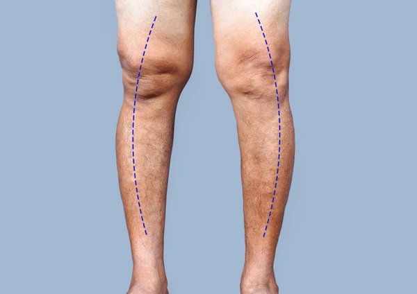
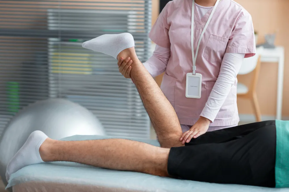
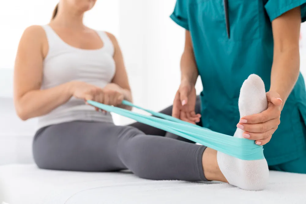
Non-surgical methods like physiotherapy or orthotics may help manage mild symptoms but do not correct bony deformities. Surgery is often the definitive solution for adults with moderate to severe bow legs.

Exercise improves strength and mobility, but cannot realign bones once they are fully developed. It is beneficial post-correction or as preventive care in early childhood.
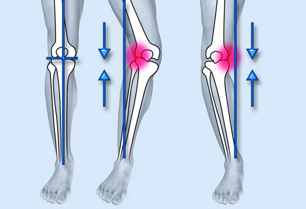
Yes, untreated bow legs can lead to arthritis, gait issues, chronic pain, and deformity progression, especially in adults.
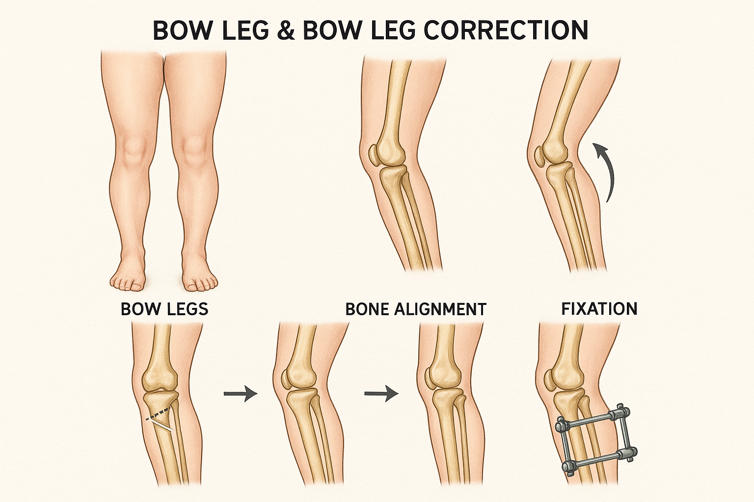
Typically between ages 4 and 11, when the growth plates are still active. Early diagnosis is key to using this minimally invasive technique.

Most insurance plans cover surgical correction when it’s medically necessary. Cosmetic procedures may not be covered unless tied to functional issues.

Most children can return within 2–3 weeks, depending on the procedure and their ability to move comfortably with crutches or walkers.

A nutrient-rich diet, consistent physiotherapy, avoiding high-impact activities, and maintaining a healthy weight are all vital for recovery and long-term joint health.