


How We Work
Frequently Asked Questions about Bow Leg Correction
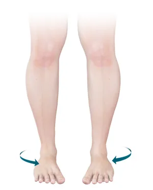
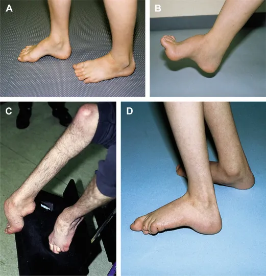
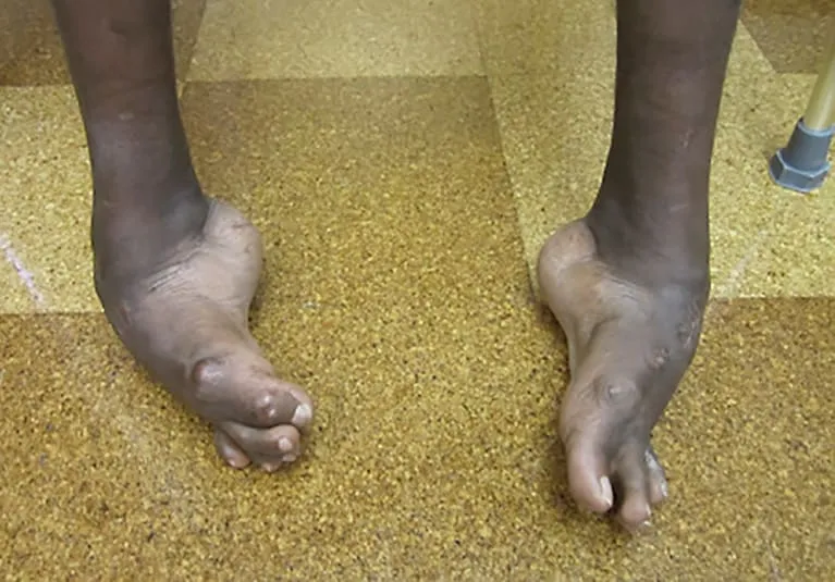
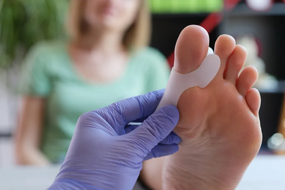
What causes foot deformities?
They can be congenital or acquired due to trauma, nerve disorders, arthritis, or poor footwear.
Can they be prevented?
Yes, through proper footwear, managing weight, and early treatment of injuries or metabolic diseases.
Are all foot deformities treatable without surgery?
No. While many mild cases improve with orthotics and therapy, advanced deformities often need surgical correction.
What results can I expect from treatment?
Most patients experience significant pain relief, improved alignment, and better walking ability.
How long does recovery take?
Recovery ranges from weeks to months, depending on treatment type and severity of deformity.
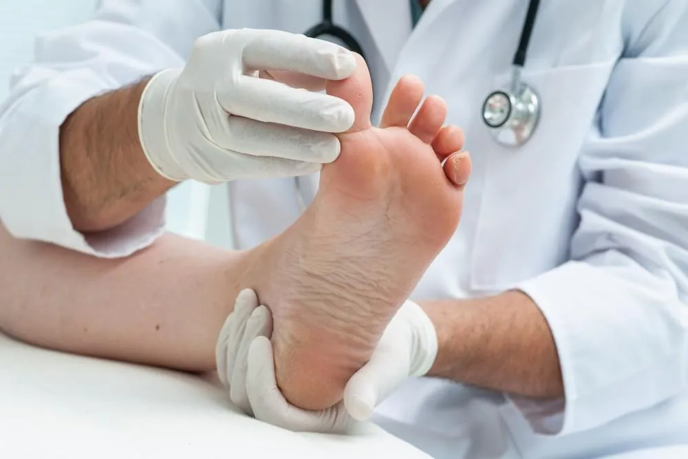
Are foot deformities painful?
They can cause pain, especially during activity or prolonged standing.
Is surgery safe for older adults?
Yes, with modern techniques and proper evaluation, elderly patients can safely undergo correction.
Can foot deformities return after treatment?
They can if the root cause isn’t addressed. Regular follow-up helps prevent recurrence.
What is the success rate for foot deformity correction?
Success rates are high, particularly when managed by experienced orthopaedic surgeons.
Will I need special shoes after treatment?
Custom footwear may be needed post-recovery to maintain correction and support.