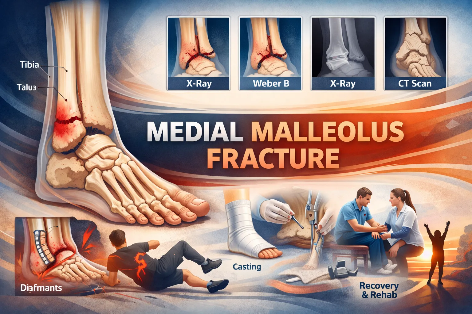
A medial malleolus fracture is a break in the inner part of the ankle that plays a key role in joint stability and weight-bearing. It commonly occurs due to twisting injuries, sports trauma, falls, or accidents, and may appear alone or with other ankle fractures. Symptoms usually include inner ankle pain, swelling, bruising, and difficulty walking. Diagnosis is confirmed through clinical examination and X-rays, with CT scans used in complex cases. Stable fractures can often heal with casting or a walking boot, while displaced or unstable fractures may require surgical fixation to restore alignment. Recovery typically takes 6 – 12 weeks for basic healing and up to 3 – 4 months for full function, depending on severity and treatment. Early diagnosis, proper treatment, and physiotherapy are essential to ensure good recovery and prevent complications like stiffness or arthritis.
Table of Contents
What is a Medial Malleolus Fracture?
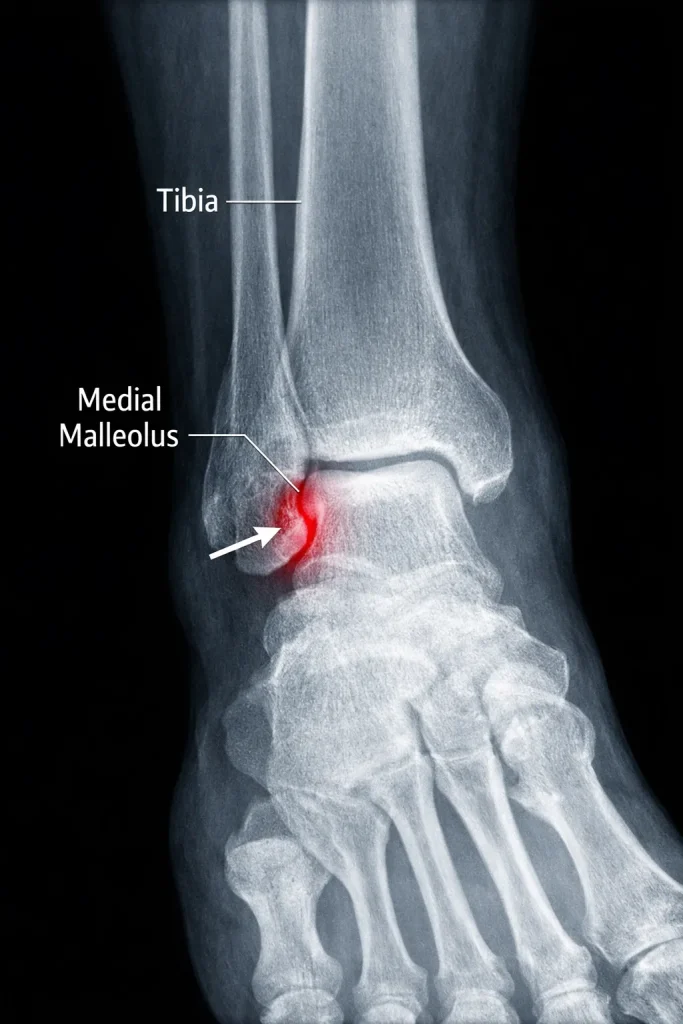
The medial malleolus is the bony prominence on the inner side of your ankle. It forms part of the ankle joint and helps stabilize the talus bone during walking and weight-bearing.
A medial malleolus fracture occurs when this inner ankle bone breaks due to twisting, impact, or a fall. It may occur:
- Alone (isolated fracture)
- With a lateral malleolus fracture (bimalleolar)
- With posterior malleolus fracture (trimalleolar)
The severity depends not only on the fracture line but also on ligament injury and joint alignment. Even a small displacement can make the ankle unstable.
Anatomy of the Medial Malleolus
The medial malleolus is part of the distal tibia and serves as an attachment for the strong deltoid ligament, which stabilizes the ankle joint.
It plays an important role in:
- Supporting body weight during walking
- Preventing lateral shift of the talus
- Maintaining ankle alignment
- Providing ligament stability
Because this structure is crucial for joint stability, improper healing can lead to long-term pain and arthritis.
Causes of Medial Malleolus Fracture
Medial malleolus fractures usually result from twisting injuries or direct trauma to the ankle. They are common in sports injuries, falls, and accidents.
Common causes include:
- Sudden twisting while walking or running
- Sports injuries (football, basketball, running)
- Slip and fall accidents
- Road traffic accidents
- Fall from height
- Osteoporosis-related fragility fractures in the elderly
In younger individuals, high-energy trauma is often responsible, while in older adults, minor falls can cause fractures due to weaker bones.
Types of Medial Malleolus Fractures
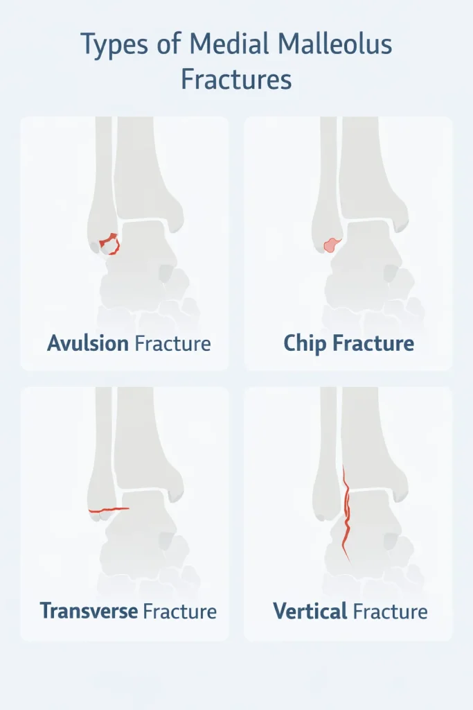
Not all medial malleolus fractures are the same. The fracture pattern determines treatment and stability.
Avulsion Fracture
An avulsion fracture occurs when a small piece of bone is pulled off by the attached ligament. These are often small and may be stable if the displacement is minimal.
Chip Fracture
A chip fracture involves a small bone fragment breaking off. These may sometimes be treated conservatively if alignment is maintained.
Transverse Fracture
A horizontal fracture line across the medial malleolus. These fractures may become unstable if displaced and often require surgical fixation.
Vertical Fracture
A vertical fracture is usually caused by high-energy injury and may extend into the ankle joint. These are more unstable and frequently require surgery.
Understanding the fracture pattern helps determine whether casting or surgery is necessary.
Symptoms of Medial Malleolus Fracture
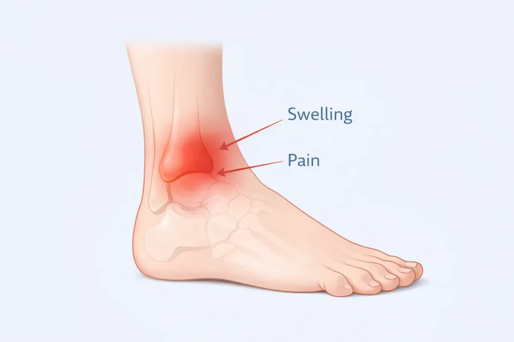
Symptoms typically appear immediately after injury but may vary depending on severity.
Common symptoms include:
- Pain on the inner side of the ankle
- Swelling around the ankle joint
- Bruising or discoloration
- Difficulty bearing weight
- Tenderness when touching the inner ankle
- Reduced range of motion
In severe fractures, visible deformity or inability to stand may occur. Persistent pain after an ankle injury should always be evaluated with imaging.
Diagnosis
Accurate diagnosis ensures proper treatment and prevents complications.
Clinical Examination
An orthopedic specialist evaluates:
- Swelling
- Bone tenderness
- Deformity
- Stability of joint
- Neurovascular status
X-ray Imaging
X-rays are the primary diagnostic tool. Standard views include:
- AP view
- Lateral view
- Mortise view
These help determine fracture pattern and displacement.
CT Scan
A CT scan may be recommended when:
- Fracture extends into the joint
- Multiple fragments present
- Surgical planning required
Advanced imaging helps assess stability and guide treatment.
Is a Medial Malleolus Fracture Stable or Unstable?
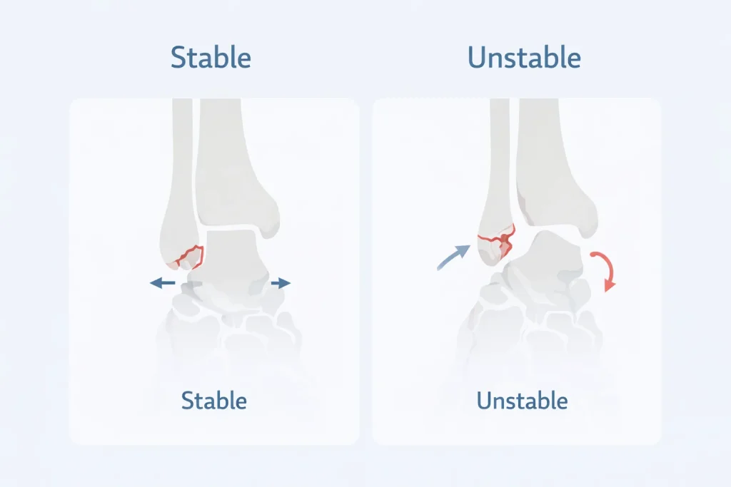
This is the most important question in treatment planning.
A fracture is considered stable if:
- Bone alignment is maintained
- No talar shift
- Ligaments intact
A fracture is unstable if:
- The bone is displaced
- Associated ligament injury present
- Part of a bimalleolar or trimalleolar fracture
Stable fractures can heal with casting, while unstable fractures usually require surgical fixation.
Treatment of Medial Malleolus Fracture
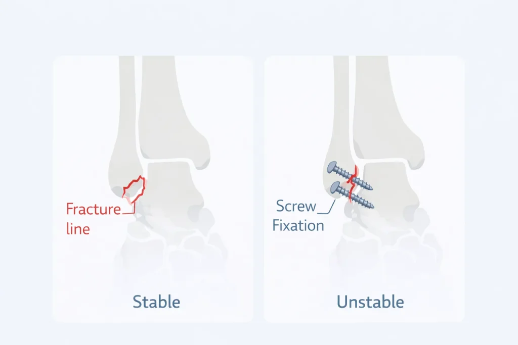
Treatment depends on:
- Displacement
- Stability
- Patient age
- Activity level
- Associated injuries
Non-Surgical Treatment
Stable fractures without displacement may heal with conservative care.
Treatment includes:
- Short leg cast or walking boot
- Non-weight bearing for 4 – 6 weeks
- Pain control
- Follow-up X-rays
- Gradual physiotherapy
Many isolated, stable fractures heal successfully without surgery.
Surgical Treatment (ORIF)
Surgery is required when:
- Fracture is displaced
- Joint unstable
- Associated fractures present
- Ligament injury exists
The procedure involves:
- Realigning bone
- Fixation with screws or a plate
- Restoring joint alignment
Surgical fixation helps restore ankle stability and reduces the risk of arthritis.
Dr Divya Ahuja performs advanced fixation techniques focused on anatomical alignment and early rehabilitation for optimal recovery.
Recovery Time After Medial Malleolus Fracture
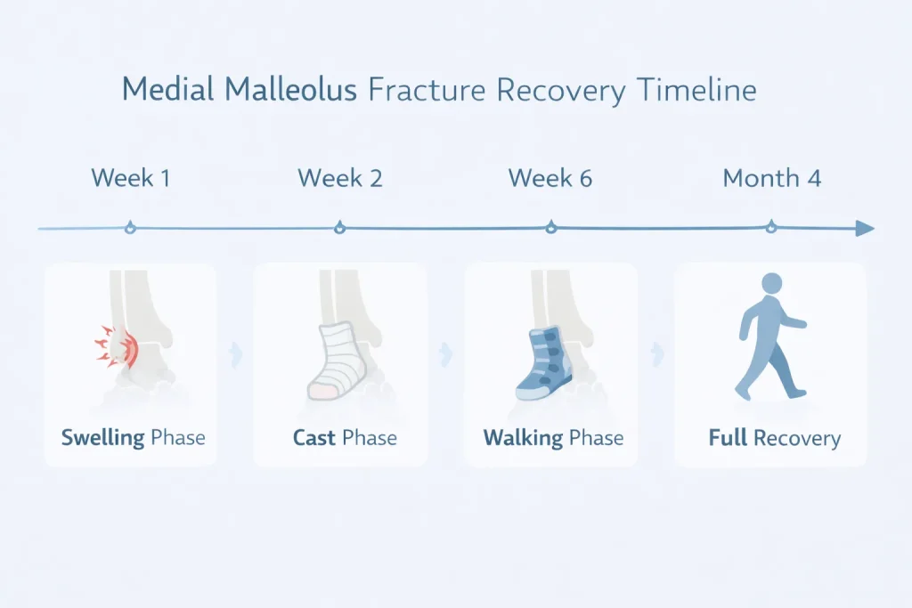
Recovery varies depending on severity and treatment.
| Treatment Type | Weight Bearing | Full Recovery |
|---|---|---|
| Stable fracture (cast) | 4 – 6 weeks | 8 – 10 weeks |
| Surgical fixation | 6 – 8 weeks | 3 – 4 months |
| Complex fracture | 8 – 10 weeks | 4 – 6 months |
Most patients regain good function with proper rehabilitation.
Recovery phases include:
- Swelling reduction phase
- Bone healing phase
- Strength rebuilding
- Balance training
- Return to activity
Swelling may persist for months even after bone healing.
Physiotherapy & Rehabilitation
Rehabilitation is crucial for restoring mobility and preventing stiffness.
Therapy includes:
- Range of motion exercises
- Strength training
- Balance exercises
- Gait correction
- Return-to-sport training
Skipping physiotherapy may result in stiffness and weakness.
Complications
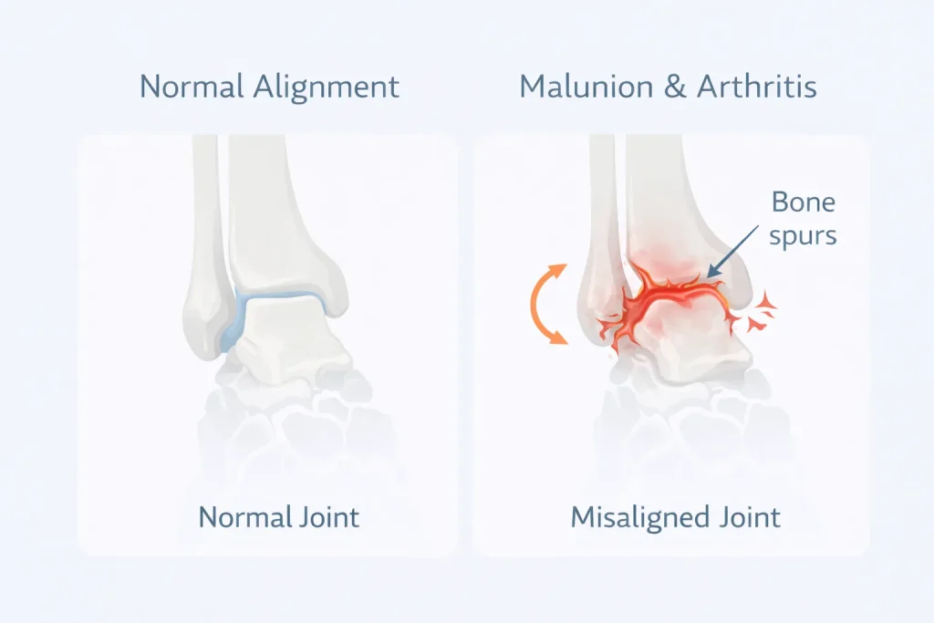
If not treated properly, complications may occur.
Possible complications include:
- Malunion
- Nonunion
- Chronic pain
- Arthritis
- Stiffness
- Hardware irritation
Early treatment and proper alignment significantly reduce risks.
When to See an Orthopedic Specialist
Seek expert care if:
- Severe ankle pain persists
- Swelling increases
- Cannot bear weight
- Deformity visible
- Injury followed by a twist or a fall
Early evaluation improves outcomes and prevents chronic problems.
Treatment in India & International Consultation
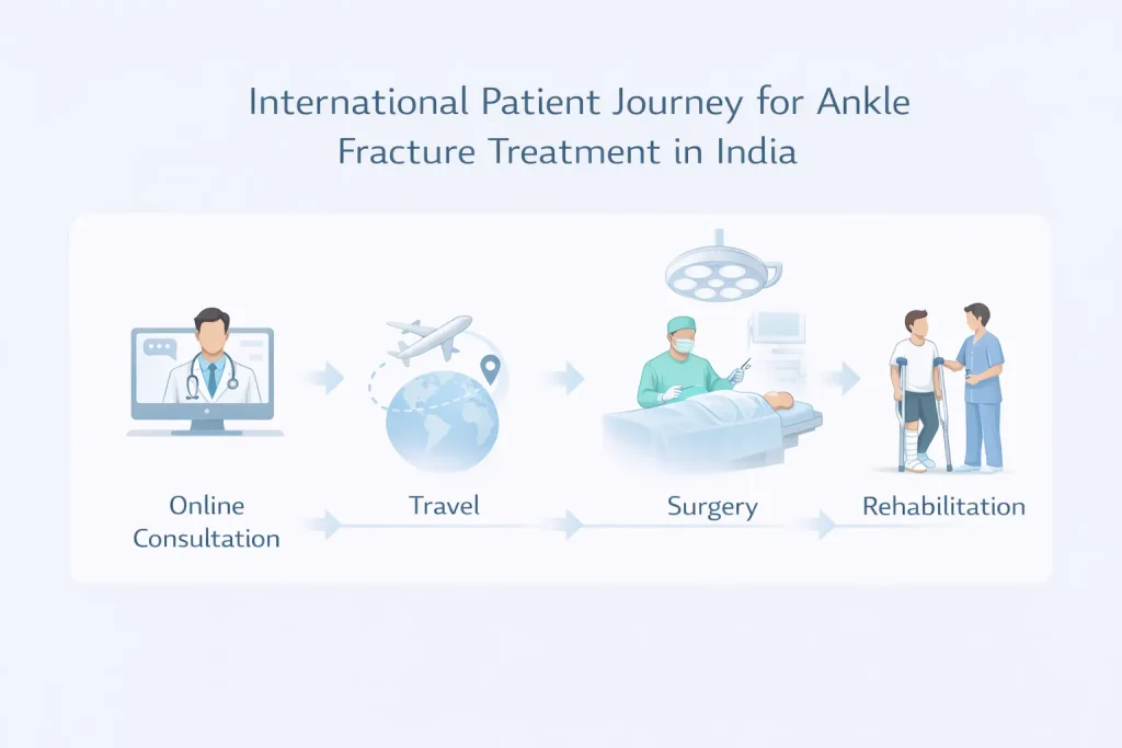
Many global patients seek orthopedic treatment in India due to advanced surgical care and cost advantages.
Dr Divya Ahuja provides specialized care for:
- Medial malleolus fractures
- Complex ankle injuries
- Revision surgeries
- Sports trauma
International patients benefit from:
- Online consultation
- Treatment planning
- Affordable surgery
- Rehabilitation guidance
Early expert treatment ensures proper healing, stable joint alignment, and faster return to activity.
Conclusion
A medial malleolus fracture is more than just a small bone injury — it directly affects ankle stability and long-term function. With timely diagnosis, proper treatment, and structured rehabilitation, most patients recover well and return to normal activities.
If you suspect an ankle fracture, early orthopedic evaluation ensures the best outcome and prevents long-term complications.
Explore all our fracture-related blogs
| Topic | Link |
| Cervical Radiculopathy | Click here |
| Distal Radius Fracture | Click here |
| Proximal Humerus Fracture | Click here |
| Lauge-Hansen Classification of Ankle Fractures | Click here |
| Weber Classification of Ankle Fractures | Click here |
| Trimalleolar Fracture | Click here |
| Bimalleolar Ankle Fracture | Click here |
| Lateral Malleolus Fracture | Click here |
| Medial Malleolus Fracture | Click here |
| Complete Guide to Ankle Fractures | Click here |
| Femur Shaft Fractures | Click here |
| Tibia Fracture | Click here |
FAQs
How long does a medial malleolus fracture take to heal?
Most medial malleolus fractures heal in 6 – 8 weeks if stable. Surgical cases may require 3 – 4 months for full recovery, including physiotherapy. Healing time varies based on fracture severity, patient age, and adherence to rehabilitation protocols.
Can you walk with a medial malleolus fracture?
Walking with a medial malleolus fracture depends on stability. Stable fractures may allow limited weight-bearing in a boot, but unstable fractures require immobilization or surgery. Walking too early can worsen displacement and delay healing.
Does a medial malleolus fracture always need surgery?
Not all medial malleolus fractures need surgery. Stable, non-displaced fractures can heal with casting. Surgery is recommended when there is displacement, instability, or associated ankle fractures to restore joint alignment.
What is a medial malleolus avulsion fracture?
An avulsion fracture occurs when a ligament pulls off a small piece of bone from the medial malleolus. These fractures may be stable and treated conservatively if alignment is maintained, but larger fragments may require fixation.
How long does swelling last after a medial malleolus fracture?
Swelling usually improves within 2 – 3 weeks, but mild swelling can persist for several months. Elevation, compression, and physiotherapy help reduce swelling and improve circulation during recovery.
What happens if a medial malleolus fracture is untreated?
Untreated fractures can heal improperly, leading to ankle instability, chronic pain, and arthritis. Proper alignment and treatment are essential to maintain joint function.