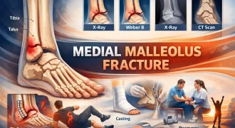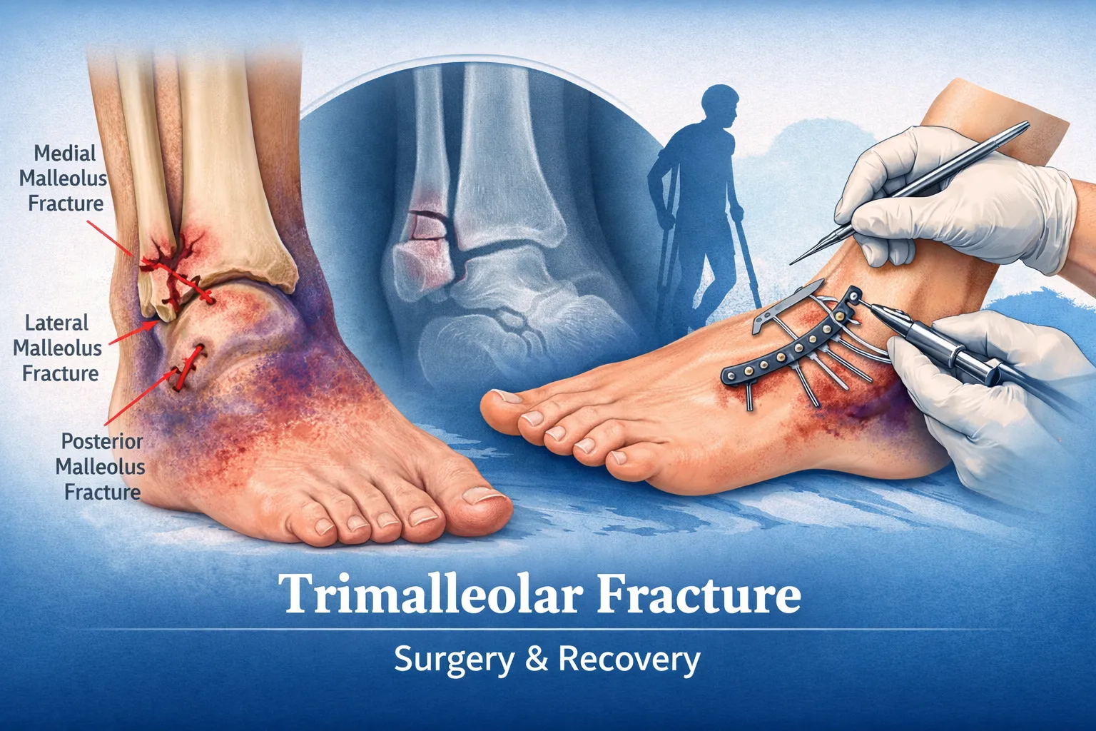
A trimalleolar fracture is a severe ankle injury involving three components of the ankle joint – the medial malleolus, lateral malleolus, and posterior malleolus. Because all major stabilizing structures are disrupted, this injury is highly unstable and almost always requires surgical fixation to restore alignment and prevent long-term complications such as arthritis, stiffness, or chronic pain.
This comprehensive guide provides in-depth explanations of trimalleolar ankle fractures, including anatomy, radiological findings, surgical techniques, fixation methods, recovery timelines, and long-term outcomes. The content is designed for patients, caregivers, medical learners, and international readers seeking expert treatment options.
Table of Contents
What is a Trimalleolar Fracture?
A trimalleolar fracture occurs when three bony structures of the ankle joint break simultaneously:
- Medial malleolus (inner ankle – tibia)
- Lateral malleolus (outer ankle – fibula)
- Posterior malleolus (back part of the distal tibia)
This injury affects the entire ankle mortise, the socket that holds the talus in place. When all three malleoli are fractured, the ankle joint becomes unstable, often accompanied by ligament injury and sometimes ankle dislocation.
Key characteristics:
- Involves three fracture components
- Highly unstable injury
- Frequently associated with ligament damage
- Almost always requires surgery
Because the posterior malleolus is involved, joint surface integrity is compromised, increasing the risk of arthritis if not properly treated.
Anatomy of the Ankle in Trimalleolar Fracture
The ankle joint consists of:
- Tibia
- Fibula
- Talus
- Medial malleolus
- Lateral malleolus
- Posterior malleolus
Together, these form the ankle mortise – a stable socket that supports body weight and allows motion.
In trimalleolar fractures:
- Both sides of the ankle break
- The posterior tibial fragment detaches
- Syndesmotic ligaments may be injured
- The talus may shift out of alignment
Because all structural supports are compromised, joint congruity must be restored surgically.
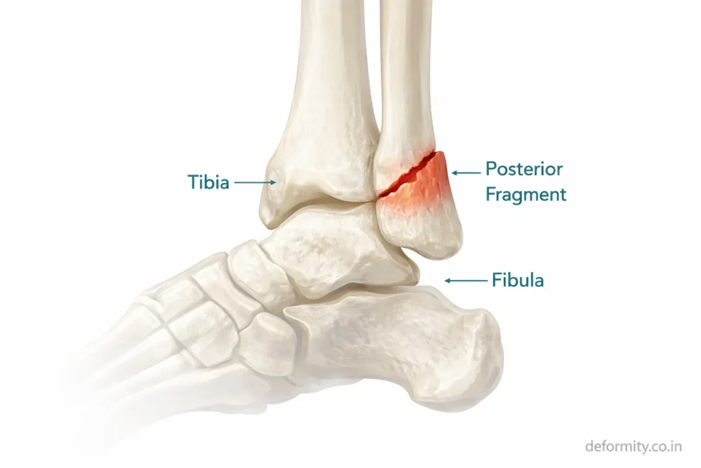
Why the Posterior Malleolus Matters
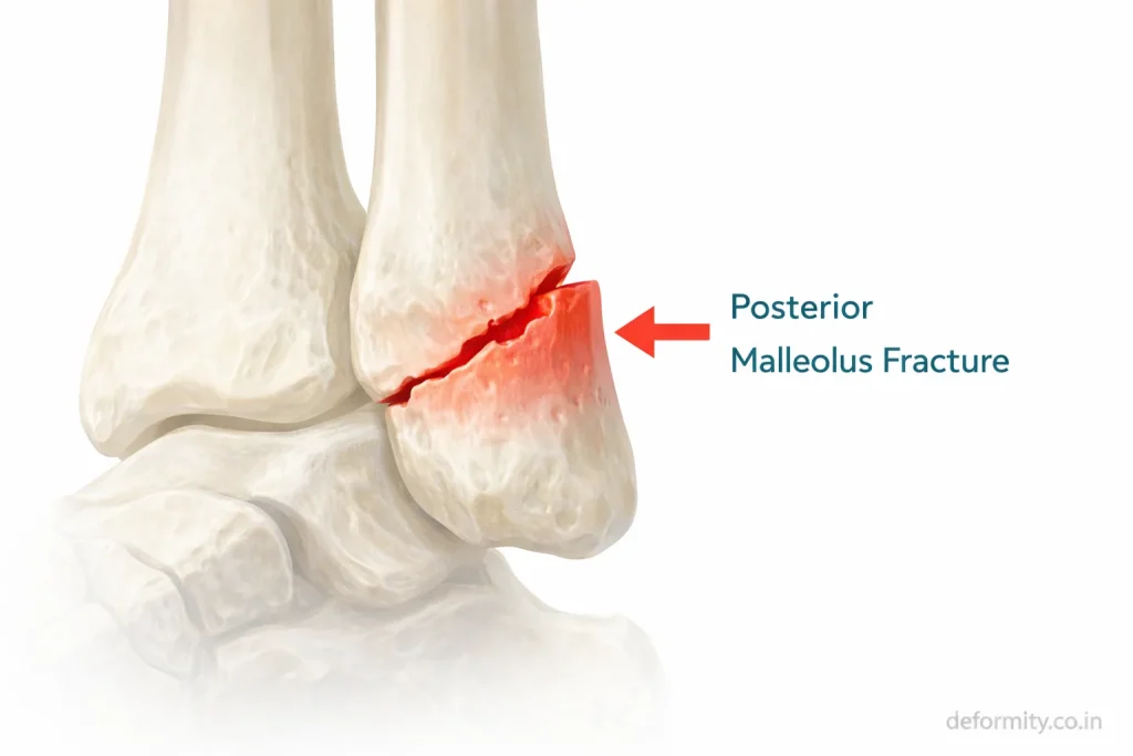
The posterior malleolus is the back portion of the distal tibia. It forms part of the weight-bearing surface of the ankle joint and provides attachment to the posterior inferior tibiofibular ligament.
Its importance includes:
- Maintaining ankle stability
- Supporting the joint surface
- Preserving syndesmotic integrity
- Preventing talar displacement
When the posterior fragment involves more than 25–30% of the joint surface, surgical fixation is strongly recommended to restore alignment and reduce the risk of arthritis.
Causes of Trimalleolar Ankle Fracture
Trimalleolar fractures usually result from high-energy trauma or severe rotational forces.
Common causes include:
- Road traffic accidents
- Fall from height
- Severe ankle twisting
- Sports trauma
- High-impact injuries
- Osteoporotic fractures in the elderly
Young individuals typically sustain these injuries in accidents, while older adults may suffer them after falls due to bone fragility.
Mechanism of Injury
Most trimalleolar fractures occur due to rotational forces applied to the ankle.
Typical mechanisms:
- Supination-external rotation injury
- Pronation injury
- Twisting with planted foot
- High rotational trauma
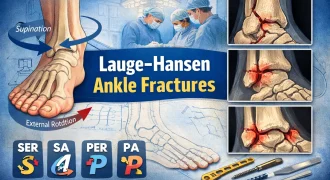
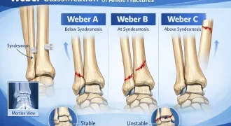
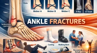
Classification systems such as Weber and Lauge-Hansen help describe fracture patterns and guide surgical planning.
Symptoms of Trimalleolar Fracture
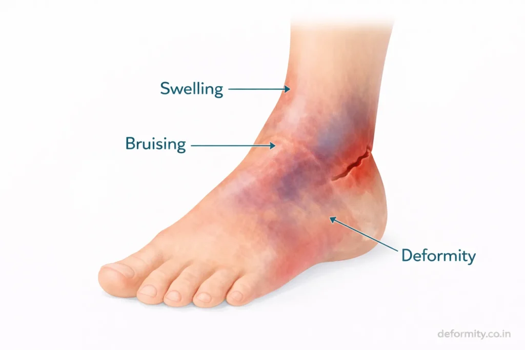
Symptoms are typically severe and immediate.
Common signs include:
- Intense ankle pain
- Rapid swelling
- Bruising
- Visible deformity
- Inability to bear weight
- Restricted movement
Because the joint is unstable, walking is usually impossible without medical support.
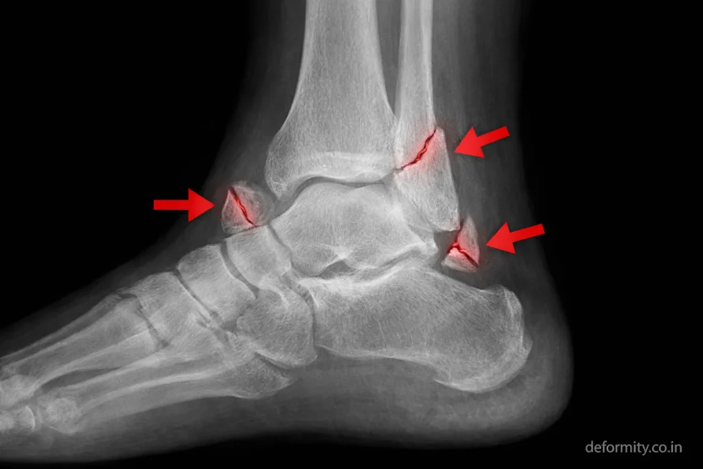
Trimalleolar Fracture Radiology
Radiology plays a critical role in diagnosis and surgical planning.
X-ray Findings
Standard X-ray views include:
- AP view
- Lateral view
- Mortise view
Findings may show:
- Fracture lines on the medial and lateral sides
- Posterior malleolus fragment on lateral view
- Joint misalignment
- Talar shift
CT Scan Importance
CT scans are often recommended because:
- Posterior fragment size must be measured
- Joint surface congruity must be assessed
- Fragment displacement must be evaluated
- Surgical planning requires precise imaging
CT imaging provides a 3D understanding of fracture geometry and helps determine fixation strategy.
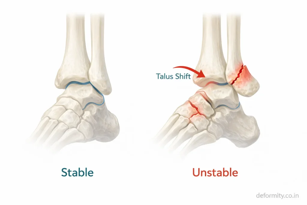
Radiology Features of Instability
Radiological signs of instability include:
- Syndesmotic widening
- Medial clear space widening
- Talar displacement
- Posterior fragment displacement
Detailed radiological evaluation ensures accurate treatment planning.
Is a Trimalleolar Fracture Always Surgical?
In most cases, yes.
Because three stabilizing structures are involved, trimalleolar fractures are generally unstable and require surgical fixation. Non-surgical treatment may be considered only if:
- Fractures are non-displaced
- Patient is medically unfit for surgery
- Functional demand is very low
However, surgical treatment typically offers better long-term outcomes.
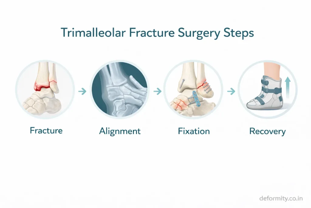
Surgical Treatment of Trimalleolar Fracture
Surgery aims to restore anatomical alignment and joint congruity.
ORIF (Open Reduction and Internal Fixation)
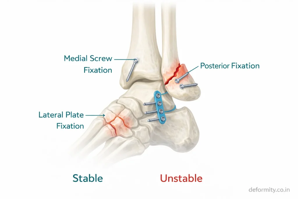
The procedure involves:
- Realigning fracture fragments
- Fixing the medial malleolus with screws
- Fixing the lateral malleolus with a plate and screws
- Fixing the posterior malleolus with screws or a plate
Posterior Malleolus Fixation Techniques
Common approaches include:
- Posterolateral approach
- Anterior-to-posterior screw fixation
- Posterior plating
Proper fixation restores joint stability and prevents talar shift.
Syndesmotic Fixation
If the syndesmotic ligaments are injured:
- Syndesmotic screws
- Tightrope fixation
may be used to maintain alignment during healing.
Dr Divya Ahuja specializes in complex ankle trauma fixation with emphasis on anatomical restoration and early rehabilitation.
Recovery After Trimalleolar Fracture
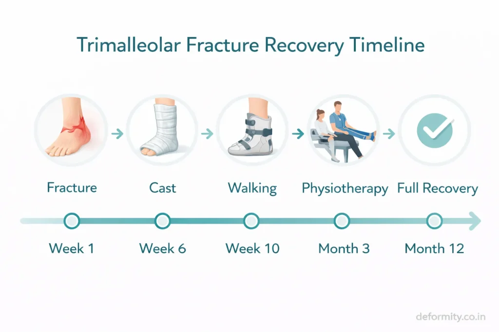
Recovery is longer compared to simpler ankle fractures.
| Stage | Timeline |
|---|---|
| Non-weight bearing | 6 weeks |
| Partial weight-bearing | 8–10 weeks |
| Walking without support | 10–12 weeks |
| Functional recovery | 3–6 months |
| Full recovery | 6–12 months |
Factors influencing recovery:
- Age
- Bone quality
- Rehabilitation adherence
- Severity of fracture
- Accuracy of fixation
Swelling may persist for several months even after bone healing.
Rehabilitation & Physiotherapy
Rehabilitation is essential for restoring function.
Key components:
- Range of motion exercises
- Strength training
- Proprioception training
- Balance exercises
- Gait correction
- Return-to-sport training
Delayed rehabilitation may result in stiffness and weakness.
Complications of Trimalleolar Fracture
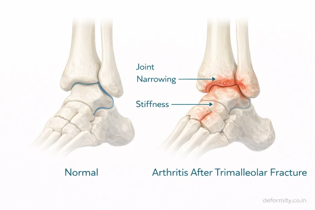
Because this injury involves joint surface disruption, complications may occur.
Possible complications:
- Post-traumatic arthritis
- Malunion
- Nonunion
- Infection
- Hardware irritation
- Chronic pain
- Reduced ankle mobility
Early surgery and structured rehabilitation significantly reduce these risks.
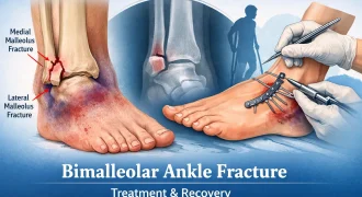
Trimalleolar vs Bimalleolar Fracture
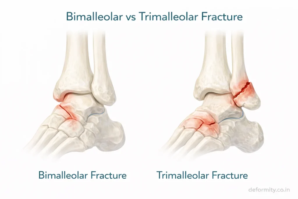
| Feature | Bimalleolar | Trimalleolar |
|---|---|---|
| Bones involved | 2 | 3 |
| Posterior involvement | No | Yes |
| Stability | Unstable | Highly unstable |
| Surgical complexity | Moderate | High |
| Recovery time | 3–6 months | 6–12 months |
Trimalleolar fractures carry a higher risk of arthritis due to posterior joint involvement.
Read more about: Bimalleolar Ankle Fracture: Symptoms, Surgery, Fixation & Recovery
When to Seek Emergency Care
Seek urgent medical attention if:
- Severe ankle deformity
- Intense swelling
- Numbness in toes
- Inability to move the foot
- Severe pain after the accident
Prompt evaluation prevents complications.
Treatment in India & International Consultation
Advanced surgical management is essential for optimal outcomes.
Dr. Divya Ahuja provides specialized treatment for complex ankle injuries, including trimalleolar fractures, witha focus on:
- Anatomical reduction
- Advanced fixation techniques
- Syndesmotic stabilization
- Structured rehabilitation
International patients benefit from:
- Online consultation
- Treatment planning
- Affordable surgery
- Comprehensive post-operative care
Conclusion
A trimalleolar fracture is a complex and unstable ankle injury requiring careful radiological evaluation, surgical fixation, and structured rehabilitation. With timely expert management and consistent physiotherapy, most patients regain functional mobility and return to daily activities. Early treatment is critical to preserve joint health and prevent long-term complications.
Read: Ankle Fractures Explained: Types, Classification, Treatment & Recovery Guide
FAQs
What is a trimalleolar fracture?
A trimalleolar fracture is a severe ankle injury involving the medial, lateral, and posterior malleoli. It disrupts joint stability and usually requires surgical fixation to restore alignment and prevent long-term complications.
Is a trimalleolar fracture serious?
Yes. Because three parts of the ankle joint are fractured, the injury is highly unstable and requires urgent orthopedic care to prevent chronic pain and arthritis.
Does it always need surgery?
Most trimalleolar fractures require surgery due to instability and joint surface involvement. Rare non-displaced cases may be managed conservatively under close supervision.
How long does trimalleolar fracture recovery take?
Basic healing occurs in 8–12 weeks, but full recovery, including strength and mobility, may take 6–12 months depending on rehabilitation and fracture severity.
What are the radiology findings in a trimalleolar fracture?
Radiology shows fractures of the medial and lateral malleoli along with a posterior tibial fragment. A CT scan helps assess fragment size and joint congruity for surgical planning.