A tibia fracture – commonly called a shinbone fracture or leg bone fracture – is one of the most serious and frequent injuries seen in orthopedic practice. The tibia is the larger of the two lower leg bones, sitting between the knee and the ankle. It carries most of your body weight with every step, which is why when it breaks, the impact on daily life is significant.
Whether caused by a high-speed road accident, a sports collision, or a fall, a broken tibia demands expert medical attention. This guide covers everything you need to know – from understanding the anatomy of the tibia and fibula, to recognizing tibia fracture symptoms, exploring treatment options including tibia rod surgery, and knowing what to expect during recovery.
This article is written for patients recently diagnosed with a tibia fracture, caregivers supporting recovery, and anyone who wants to understand what a tibia fracture really means for the body.
Table of Contents
What Is the Tibia? Understanding the Shin Bone
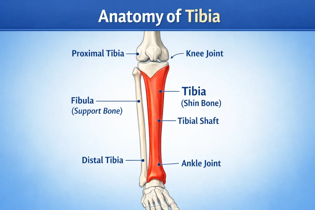
Before discussing fractures, it helps to understand the bone itself. The tibia – also called the shin bone – is the larger and stronger of the two lower leg bones. Located between the ankle and the knee, it forms a critical part of both the knee joint above and the ankle joint below.
The tibia and fibula together make up the bony framework of the lower leg. While the tibia is weight-bearing and structural, the fibula (the thinner bone alongside it) mainly provides stability and anchors muscles. This is why in many tibia fractures, the fibula breaks at the same time.
Key facts about the tibia bone:
- The tibia is the most commonly fractured long bone in the entire human body
- It is the shin body part you can feel by pressing your lower leg – the hard ridge running down the front
- It supports the majority of body weight during standing and walking
- The tibia has three anatomical sections: proximal (upper end), tibial shaft (middle), and distal (lower end of tibia)
- The shin bone, named in medical terminology, is the tibia, derived from Latin, meaning flute, referring to its shape
- The inner ankle bone (medial malleolus) is actually part of the distal end of the tibia
- The bone between the ankle and knee is the tibia – a common question from patients trying to understand their anatomy
The tibial shaft refers specifically to the long middle section of the bone. Tibial shaft fractures occur below the knee and above the ankle, making this the most clinically relevant zone for most patients.
Is the Tibia Bone Hollow?
Yes. Like most long bones in the body, the tibia has a hollow central canal called the medullary canal. This canal is filled with bone marrow and is precisely what makes intramedullary nailing (tibia rod surgery) possible – the metal rod is inserted directly into this hollow space to stabilize the fracture.
The hollow architecture makes the tibia strong under normal loads while remaining lightweight. However, it also means that when a fracture occurs, the bone can shatter into multiple fragments under high-energy impact – producing the comminuted fractures commonly seen in road traffic accidents.
How Strong Is the Tibia?
The tibia is one of the strongest bones in the human body. It can withstand forces of approximately 4,000 to 7,000 Newtons during normal walking – roughly 4 to 7 times body weight. This strength is why it typically takes significant trauma to fracture a healthy tibia in a young adult.
However, bone strength decreases with age, osteoporosis, vitamin D deficiency, and certain medications. In elderly patients, relatively minor falls can produce significant tibia fractures because the bone is no longer at full structural strength.
What is a Tibia Fracture?
A tibia fracture means the continuity of the tibia bone has been broken, partially or completely. A tibial shaft fracture is a break occurring along the length of the bone between the knee and ankle joints. These are among the most common leg bone fractures treated in orthopedic hospitals across India and worldwide.
In many cases, the fibula is also broken alongside the tibia. This is called a both-bone leg fracture or a tib-fib fracture. Severity can range from a hairline fracture – a thin crack in the bone – to a compound fracture of the tibia where the bone pierces through the skin.
What is a tibia fracture in simple terms?
It is a break in your shin bone that can happen with varying levels of severity, and it almost always requires professional orthopedic evaluation and treatment.
BB Leg Fracture: What Does It Mean?
Many patients in India search for “BB leg fracture” or “fracture BB leg full form”. BB stands for Both Bone – meaning both the tibia and fibula of the lower leg are fractured simultaneously.
A bilateral bone leg fracture (BB fracture leg) is more serious than a single bone fracture because:
- Both weight-bearing and stabilizing structures of the lower leg are compromised
- The leg is significantly more unstable
- Soft tissue damage is usually more extensive
- Surgery is more commonly required
Common search terms for this injury include:
- BB leg fracture
- Both bone leg fractures
- The left leg has both a bone fracture
- Right leg has both bone fractures
- Fracture both bone leg
Both bone leg fracture classification helps surgeons determine treatment – whether casting, nailing, or plating – based on the pattern and displacement of both fractures.
Types of Tibia Fractures: A Complete Classification

Not all tibia fractures are the same. Orthopedic surgeons classify them based on pattern, location, severity, and whether the skin remains intact. Understanding the type of fracture determines the correct treatment approach.
By Fracture Pattern
- Transverse fracture – A straight, horizontal break across the tibial shaft. Generally clean and often stable.
- Oblique fracture – An angled break across the shaft, typically caused by a diagonal force.
- Spiral fracture tibia – The fracture line spirals around the shaft like a corkscrew. Caused by a twisting force. Common in sports injuries.
- Comminuted fracture tibia – The bone shatters into three or more fragments. Typically caused by high-energy trauma such as road accidents. Comminuted fracture images on X-ray show a bone broken into multiple pieces.
- Segmental tibia fracture – The bone breaks in two separate places, leaving a floating middle segment of bone.
By Skin Integrity
- Closed fracture – The skin remains intact over the fracture site. Lower risk of infection. This is what is referred to as a closed surgery approach in conservative management.
- Open tibia fracture (compound fracture) – Bone fragments break through the skin, or a wound reaches the bone. High risk of infection. Requires emergency surgical treatment. A compound tibia fracture is one of the most serious presentations in orthopedics.
By Location on the Tibia Bone
- Proximal tibia fracture – Near the knee. Can involve the tibial condyle fracture or tibial spine fracture.
- Tibial shaft fracture – The mid-section of the bone. The most common type.
- Distal tibia fracture / Fracture of the lower end of the tibia – Near the ankle, sometimes involving the ankle joint. Also called a lower tibia fracture or fracture of the lower end of the tibia.
- Distal 1/3 tibia fracture – Affects the lower third of the shaft, near the ankle.
- Tibia ankle fracture – When the fracture involves both the lower tibia and the ankle joint, also described as a broken tibia ankle.
Tibial Shaft Fracture Classification
Surgeons use standardized systems – including the AO/OTA Classification and the Gustilo-Anderson Classification for open fractures – to guide treatment. The tibial shaft fracture classification and tibia shaft fracture classification systems help surgeons communicate precisely about fracture severity, location, and the degree of soft tissue involvement.
Causes of Tibia Fractures: How Does the Shinbone Break?
The tibia is a strong bone. It takes significant force to fracture it in a healthy adult. Causes generally fall into three categories.
High-Energy Trauma
- Motor vehicle and motorcycle accidents – the leading cause of tibial shaft fractures in India
- Falls from significant height
- Direct blunt force impact to the lower leg
- These tend to produce comminuted fractures and are more likely to cause open tibia fractures with damage to surrounding muscles, nerves, and blood vessels
Low-Energy and Sports Injuries
- Falls while skiing, playing football, or soccer
- Collision with another player
- Twisting injuries – a very common cause of spiral fracture tibia
- Sports-related fractures are generally less severe but still require proper orthopedic management
Stress Fractures
Tibia stress fracture symptoms develop gradually – unlike sudden traumatic fractures. They result from repetitive loading of the bone without adequate rest. Common in long-distance runners, military recruits, and dancers.
Tibia stress fracture symptoms include:
- Dull aching pain along the shin bone that worsens with physical activity
- Pain that improves with rest but returns when activity resumes
- Tenderness to touch along a specific point on the tibia
- Mild swelling over the shin
Can You Fracture Your Tibia and Still Walk?
This is one of the most frequently searched questions about tibia injuries.
The answer depends on the type and severity of the fracture:
- Hairline or stress fractures: Some patients can bear weight and walk with pain. This is actually why stress fractures are often missed – the ability to walk leads patients to believe it is “just a bruise.”
- Simple, non-displaced closed fractures: Occasionally, partial weight bearing is possible, but it is painful and risky.
- Displaced, comminuted, or open fractures: Walking is generally not possible. The leg cannot support weight, and attempting to do so risks worsening the fracture and damaging blood vessels and nerves.
Important: The ability to walk does not rule out a fracture. If you have shin pain after trauma, always get an X-ray, even if you can put some weight on the leg.
Tibia Fracture Symptoms: What Does a Broken Tibia Feel Like?

Recognizing tibia fracture symptoms quickly is critical. Most tibial shaft fractures produce immediate and unmistakable signs.
Primary tibia fracture symptoms include:
- Severe and immediate pain at the fracture site in the lower leg
- Inability to walk or bear weight on the leg
- Visible deformity – the leg may look bent, twisted, shortened, or abnormally angled
- Swelling that develops rapidly at the fracture site, sometimes extending to the foot (called tibia fracture swelling foot)
- Bruising and skin discoloration over the shinbone area
- Bone tenting – where a bone fragment pushes sharply against the skin from the inside
- In open fractures: bone is visibly protruding through broken skin
- Occasional numbness or loss of feeling in the foot (nerve involvement)
- Instability – the leg may feel structurally unsupported
What Does a Fractured Tibia Feel Like?
Patients describe the moment of a tibia fracture in very consistent ways:
- A sudden, severe crack or snap was felt in the lower leg
- Immediate, overwhelming pain – often described as the worst pain they have ever felt
- A sensation that the leg “completely gave way” or felt structurally absent
- In high-energy fractures, some patients describe a momentary numbness from shock before the pain sets in fully
After the initial moment:
- Tibia fracture pain settles into a constant, deep aching at the fracture site
- Any attempt to move or rotate the leg significantly worsens pain
- The leg may feel cold below the fracture if the blood supply is affected
- In open fractures, patients report seeing the bone and experiencing both intense pain and shock simultaneously
Signs of a Broken Tibia vs a Badly Bruised Shin
Many patients wonder whether their injury is a fracture or simply a very bad bruise. Here are the distinguishing signs:
| Feature | Broken Tibia | Badly Bruised Shin |
| Pain severity | Severe, immediate | Moderate, builds gradually |
| Weight bearing | Usually impossible | Often possible, even if painful |
| Deformity | May be present | Not present |
| Swelling onset | Very rapid (minutes) | Gradual (hours) |
| Bone tenderness | Pinpoint, severe | Diffuse, less specific |
| Bruising pattern | Often deep and widespread | More superficial |
| X-ray | Fracture line visible | Normal bone |
If in any doubt – get an X-ray. A missed tibia fracture that continues to be loaded can worsen significantly.
Shin Fracture Swelling – What to Expect
Swelling after a tibia fracture is expected and is often significant in the first 48 to 72 hours. Shin fracture swelling can extend to the ankle and foot. This is why doctors often apply a splint initially rather than a full cast – to allow the leg to swell safely before definitive treatment is applied.
Important Warning: If you experience intense, burning pain inside the lower leg that feels completely out of proportion to the injury – especially if the leg feels extremely tight and swollen – seek emergency care immediately. This may be compartment syndrome, a serious and time-sensitive complication of tibia fractures.
Tibia Fracture Healing Stages
Understanding how a broken tibia heals helps patients follow their recovery with realistic expectations.
Stage 1 – Hematoma Formation (Days 1–5)
Immediately after a fracture, bleeding occurs around the break site. The blood clots to form a hematoma (blood pool) around the fracture. This is painful and produces significant swelling. The hematoma is not a problem – it is the essential foundation for healing.
Stage 2 – Soft Callus Formation (Weeks 1–4)
The body begins laying down soft, fibrous tissue called a soft callus around the fracture site. This callus is not yet bone – it is more like cartilage – but it begins to bridge the gap between the broken ends. Pain starts to reduce. The leg remains fragile and must be protected.
Stage 3 – Hard Callus Formation (Weeks 4–12)
The soft callus is gradually replaced by hard, calcified new bone. On the tibia fracture X-ray, this appears as a fuzzy white shadow around the fracture site – a very reassuring sign called the callus shadow. The tibia fracture after 8 weeks should show early callus formation clearly on imaging.
Stage 4 – Bone Remodeling (Months 3–12+)
The new bone gradually remodels and strengthens along the lines of mechanical stress. The callus shrinks, and the fracture line becomes less visible on X-ray. Eventually, the bone returns to near-original strength. Full remodeling can take up to 2 years in adults.
Shin Fracture Swelling – What to Expect
Swelling after a tibia fracture is expected and is often significant in the first 48 to 72 hours. Shin fracture swelling can extend to the ankle and foot. This is why doctors often apply a splint initially rather than a full cast – to allow the leg to swell safely before definitive treatment is applied.
Important Warning: If you experience intense, burning pain inside the lower leg that feels completely out of proportion to the injury – especially if the leg feels extremely tight and swollen – seek emergency care immediately. This may be compartment syndrome, a serious and time-sensitive complication of tibia fractures.
Diagnosing a Tibia Fracture: How Doctors Confirm the Injury
Medical History and Clinical Examination
Your orthopedic surgeon will begin with a detailed history – how the injury happened, the speed of impact, seatbelt use in vehicle accidents, and your overall health, including conditions like diabetes, high blood pressure, or asthma. This context helps determine fracture severity and risk of complications.
A careful physical examination follows, assessing deformity, skin integrity, swelling, bruising, and whether pulses and sensation in the foot are intact.
Imaging Tests
Diagnosis is confirmed with imaging studies.
- Tibia fracture X-ray – The most important first investigation. An X-ray of the leg shows the tibia and fibula clearly, confirming the fracture type and location. A right tibia fracture X-ray, left tibia fracture X-ray, or right leg fracture X-ray typically includes both the knee and ankle to check joint involvement. A leg X-ray is fast, widely available, and highly accurate for fracture diagnosis.
- CT Scan – Used when the tibia fracture X-ray is unclear, or when the fracture involves a joint. CT provides cross-sectional images and reveals subtle fracture lines and bone fragmentation that X-rays may miss.
- MRI – Used mainly for tibia stress fracture symptoms or when soft tissue and ligament injury needs detailed assessment.
Hairline Fracture Tibia: Diagnosis and Treatment
A hairline fracture of the tibia (also called a tibia stress fracture) is a thin, incomplete crack that often does not appear on initial X-rays. This makes diagnosis challenging and leads to delays in treatment.
Hairline fracture tibia symptoms:
- Gradual onset of shin pain that worsens with activity
- Pain that is relieved by rest but returns immediately upon resuming activity
- Point tenderness over a specific area of the tibia
- Mild swelling along the shin bone
- No sudden trauma – the pain develops over days to weeks
How is a hairline fracture tibia diagnosed?
- The initial X-ray may appear completely normal
- MRI is the gold standard and can detect stress fractures within days of onset
- A bone scan can also detect increased bone turnover at the fracture site
Hairline fracture tibia treatment:
- Rest from high-impact activity is the primary treatment
- A walking boot or cast may be used for 6–8 weeks
- Gradual return to activity guided by symptoms and imaging
- Identifying and correcting the cause (training errors, footwear, nutritional deficiency)
Hairline fracture tibia recovery time: Most heal in 6–10 weeks with adequate rest. Return to running and high-impact sport typically takes 8–12 weeks.
Tibia Fracture Treatment: From Splints to Surgery

Treatment depends on fracture type, severity, patient age, overall health, and whether the fracture is open or closed.
Nonsurgical Tibia Fracture Treatment
Not every tibia fracture requires surgery. Nonsurgical leg fracture treatment may be appropriate for:
- Stable, closed fractures with minimal bone displacement
- Patients who are poor surgical candidates due to other medical conditions
- Less active patients who can tolerate minor alignment differences in healing
Nonsurgical treatment steps:
- Initial splinting – A splint is applied first (not a full cast) to provide support while allowing the leg to swell safely. It can be loosened as needed.
- Casting – Once swelling reduces, a full cast is applied to immobilize the fracture while it heals.
- Functional bracing – After several weeks in a cast, a removable plastic brace replaces it. The brace provides protection and support, can be removed for hygiene, and allows gentle movement and physical therapy.
Surgical Tibia Fracture Treatment
Surgery is recommended for:
- Open tibia fractures with wounds needing monitoring and cleaning
- Fractures where the bone is significantly displaced
- Comminuted fractures with multiple bone fragments
- Fractures that fail to heal with nonsurgical treatment
- Certain fracture locations near the knee or ankle joint
Intramedullary Nailing – Tibia Rod Surgery
Intramedullary nailing is the most widely used surgical technique for tibial shaft fractures worldwide. Patients commonly call this tibia rod surgery or tibial nail surgery.
- A titanium metal rod (nail) is inserted inside the hollow medullary canal of the tibia bone
- The rod passes across the fracture, keeping both ends in correct alignment
- Locking screws are placed at both the top and bottom to keep the nail and bone stable
- This internal fixation device provides strong, full-length fixation and allows earlier weight bearing compared to casting
- A tibial nail X-ray or tibial nailing X-ray shows the rod running the full length of the bone – this is one of the most searched images by patients after their surgery
Intramedullary nailing is generally not recommended for children and adolescents because the growth plates in developing bones must be protected.
Plates and Screws – ORIF Tibia (Tibia Plating Surgery)
ORIF tibia (Open Reduction Internal Fixation) involves repositioning the broken bone fragments into their normal alignment and holding them together with metal plates and screws fixed to the outer surface of the bone.
Tibia plating surgery is used when:
- Intramedullary nailing is not suitable
- The fracture extends into the knee joint (proximal tibia fracture) or ankle joint (distal tibia fracture)
- Tibial condyle fractures or distal tibia fracture management situations near joints
External Fixation
In cases of severe open tibia fractures or extensive soft tissue damage, external fixation provides a stabilizing frame outside the skin. Metal pins are inserted into the bone above and below the fracture and connected by a rigid external bar. This stabilizes the bone while wounds are treated and monitored. It may be temporary (before definitive surgery) or permanent in select cases.
Tibia Fracture Healing Time in Adults: What to Realistically Expect
The most common question patients ask is how long a tibia fracture takes to heal. The honest answer depends on the fracture type, treatment used, patient age, and lifestyle factors.
General Healing Timeline
- Most tibial shaft fractures take 4 to 6 months to heal completely in adults
- Simple, stable closed fractures may heal within the lower end of this range
- Comminuted, open, or segmental fractures can take significantly longer
- Tibia bone fracture healing time is extended in tobacco users, people with diabetes, and those with poor nutritional status
Tibia Fracture Healing Time by Fracture Type
| Fracture Type | Healing Time |
| Hairline / stress fracture tibia | 6–10 weeks |
| Simple closed transverse fracture | 3–4 months |
| Spiral fracture tibia recovery time | 4–6 months |
| Comminuted tibia fracture | 5–8 months |
| Open tibia fracture recovery time | 6–12 months |
| Distal tibia fracture healing time | 4–6 months |
| Proximal tibia fracture recovery time | 4–6 months |
| Shattered tibia recovery time | 8–12 months or more |
| Tibia fracture healing time in kids | 2–3 months (faster due to active growth) |
| Tibia fracture healing time in teens | 3–4 months |
| Broken tibia and fibula recovery time adults | 4–6 months |
Tibial Rod Surgery Recovery Time
Patients who undergo tibia rod surgery (intramedullary nailing) often achieve better functional outcomes earlier than those treated with casting alone. The general tibial rod surgery recovery time follows this pathway:
- Weeks 1–2: Post-surgical pain management, wound care, anti-swelling measures
- Weeks 2–6: Partial weight bearing with crutches begins; physiotherapy starts
- Months 2–3: Progressive weight bearing as tolerated, muscle strengthening exercises
- Months 4–6: Return to normal walking; bone healing is monitored on tibia fracture X-ray
- 6–12 months: Full return to sports or heavy physical activity for younger, active patients
Tibia Fracture After 8 Weeks
Patients often ask what to expect at the tibia fracture after the 8 weeks mark. By this point, early callus (new bone formation) should be visible on X-ray. Pain should be significantly reduced. Partial weight bearing should be comfortable for most patients. If there is no visible healing on imaging at this stage, your doctor will investigate for delayed union.
Tibia Fracture After 4 Weeks
At 4 weeks post-fracture or post-surgery, what should patients expect?
- Swelling has significantly reduced compared to the first week
- Wound healing should be well advanced in surgical cases
- Pain is noticeably lower, particularly at rest
- Soft callus formation has begun – not yet visible on X-ray but progressing internally
- Most patients remain non-weight-bearing or partial weight-bearing with crutches
- Physiotherapy for muscle maintenance (quadriceps, ankle movements) should be ongoing
- A cast or boot is still required for protection
If pain is increasing at 4 weeks rather than decreasing, or if swelling is worsening, medical review is warranted.
How Many Days After Tibia Surgery Can I Walk?
This depends on the type of surgery and fracture severity:
- After intramedullary nailing: Most patients begin partial weight bearing with crutches within 2–4 weeks of surgery. The exact timeline is guided by X-ray evidence and surgeon assessment.
- After plate fixation (ORIF): Weight bearing is typically delayed slightly longer – often 4–6 weeks – because plate fixation is more sensitive to early load.
- After external fixation: Weight bearing depends on fracture complexity and soft tissue healing.
Full, unsupported weight bearing without crutches typically happens around 8–12 weeks post-surgery for most patients. Never walk without your surgeon’s clearance, regardless of how the leg feels – premature loading can bend or break hardware and displace the healing fracture.
Shin Fracture Healing Time
Tibia stress fractures heal faster – typically 6 to 12 weeks – with rest and modified activity. Traumatic tibial shaft fractures take considerably longer due to the extent of bone and soft tissue damage involved.
Tibia Fibula Fracture Recovery Time
When both the tibia and fibula are broken together, tibia fibula fracture recovery time follows a similar 4-to-6-month framework, though this can extend further depending on the severity and treatment method used.
Broken Tibia and Fibula – Titanium Rod Recovery Time
One of the most commonly searched topics is “broken tibia and fibula titanium rod recovery time” – particularly from patients who have had intramedullary nailing for a both-bone lower leg fracture.
A titanium rod (intramedullary nail) is the standard surgical treatment for both bone leg fractures with tibial shaft involvement. Here is what recovery typically looks like:
Week 1–2: Hospitalization period; pain management; wound care; leg elevated to control swelling. No weight bearing.
Week 2–6: Discharge home with crutches. Physiotherapy begins for muscle maintenance and a gentle ankle range of motion. Most patients remain partial weight bearing.
Week 6–10: Progressive weight bearing begins based on X-ray evidence of callus formation. Physiotherapy advances in strengthening.
Month 3–4: Most patients walk short distances without crutches. Strength and endurance exercises continue. Daily activities gradually resume.
Month 4–6: Return to more demanding activities. Driving (in an automatic vehicle, in the non-operated leg) may be possible by this stage in some patients.
Month 6–12: Full recovery for most patients. Return to sports and heavy physical work for younger, active individuals.
The titanium rod remains in the bone permanently in most patients. Removal is not routinely performed unless hardware causes irritation or other problems.
Recovery After Tibia Fracture: Pain, Physiotherapy & Walking Again

Pain Management
Pain after a tibia fracture or surgery is expected and manageable. Your orthopedic team will use a combination approach, including:
- Acetaminophen and NSAIDs (anti-inflammatory drugs)
- Gabapentinoids for nerve-related pain
- Muscle relaxants if spasm is significant
- Short-term opioids in severe cases – used carefully and tapered as quickly as possible
- Topical pain medications are applied directly to the skin over the fracture area
Tibia Fracture Swelling: How Long Does It Last?
Swelling is one of the most persistent and frustrating aspects of tibia fracture recovery.
- First 48–72 hours: Swelling peaks. The ankle and foot may swell significantly even when the fracture is in the mid-shaft.
- Week 1–4: Swelling gradually reduces with elevation, compression, and immobilization.
- Month 1–3: Residual swelling around the fracture site and lower leg is common, especially after activity.
- Month 3–6: Most swelling resolves, though mild puffiness around the ankle and shin can persist.
- Beyond 6 months: Persistent swelling should be evaluated – it may indicate incomplete healing, hardware issues, or early arthritis if the ankle joint was involved.
Tips to reduce tibia fracture swelling:
- Elevate the leg above heart level when resting
- Perform gentle ankle pump exercises (moving the foot up and down) to encourage fluid drainage
- Use compression stockings as advised by your physiotherapist
- Avoid prolonged standing or sitting with the leg dependent
Physiotherapy – Tibia Fracture Exercises
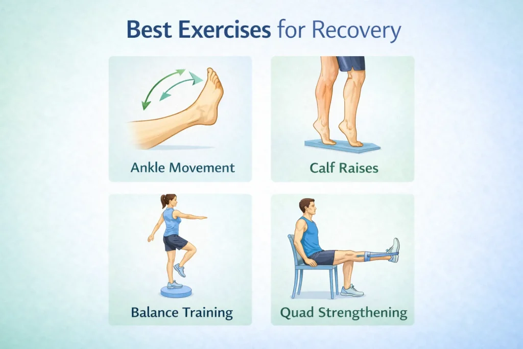
Physiotherapy is essential. Muscles weaken rapidly during immobilization, and joints stiffen. A physiotherapist will guide you through structured tibia fracture exercises:
- Quadriceps strengthening exercises to rebuild knee and thigh support
- Ankle range-of-motion exercises to prevent stiffness
- Progressive weight-bearing and gait re-training
- Balance and proprioception (body awareness) exercises
- Calf stretching and strengthening once healing allows
Walking After Broken Tibia and Fibula
Most patients need crutches or a walker for the first several weeks. Walking after broken tibia and fibula injuries is a gradual, supervised process. Your surgeon will guide weight-bearing decisions based on X-ray evidence of bone healing – not just how the leg feels. Rushing weight-bearing risks re-fracture or hardware failure.
Tibia Fracture Recovery Timeline: Month by Month
| Timeframe | What to Expect |
| Days 1–7 | Hospitalization or immobilization; pain and swelling management; no weight bearing |
| Weeks 2–4 | Wound healing; cast or splint in place; gentle physiotherapy starts |
| Tibia fracture after 4 weeks | Soft callus forming internally; pain decreasing; partial weight bearing may begin |
| Tibia fracture after 8 weeks | Hard callus visible on X-ray; progressive weight bearing; walking with support |
| Month 3–4 | Most patients walking with reduced support; active strengthening ongoing |
| Month 4–6 | Return to most daily activities; bone consolidating; driving may resume |
| Month 6–12 | Full recovery for most; return to sport and heavy work; tibia near full strength |
| Beyond 12 months | Bone remodeling continues; final strength achieved; X-ray shows healed fracture |
Complications of Tibia Fractures: What Can Go Wrong?
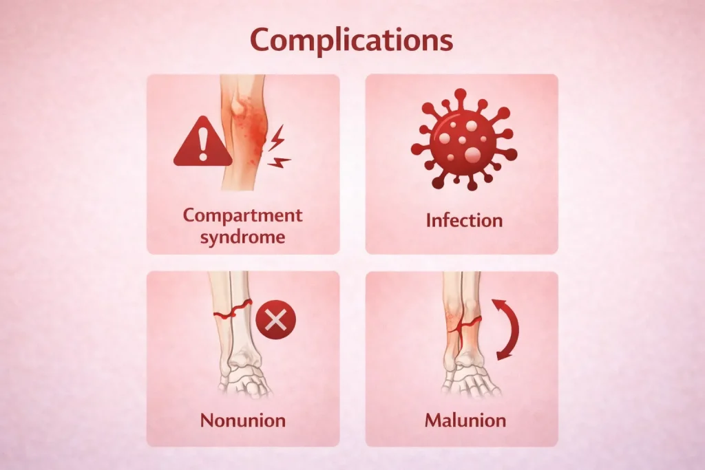
Knowing the potential complications allows patients and caregivers to recognize warning signs early and seek help promptly.
Compartment Syndrome
This is the most dangerous early complication of a tibia fracture. Swelling inside the tight muscle compartments of the lower leg builds pressure to dangerous levels, cutting off blood supply to muscles and nerves.
Symptoms include:
- Severe, burning pain inside the lower leg that feels completely out of proportion to the injury
- A tight, wooden feeling in the leg
- Numbness or tingling in the foot
- Pain that worsens when the toes are passively stretched
This is a surgical emergency. Without immediate fasciotomy (surgical release of the compartment pressure), permanent muscle and nerve damage can result.
Infection
Open tibia fractures and tibia fracture surgery both carry an infection risk. Bone infection (osteomyelitis) is particularly difficult to treat and often requires multiple surgeries and long-term antibiotics.
Nonunion and Delayed Union
If the tibia bone fracture healing time extends far beyond expected timelines without bone healing visible on X-ray, this is called delayed union. If healing stops completely, it is nonunion. Risk factors include smoking, uncontrolled diabetes, poor nutrition, and inadequate fracture fixation.
Malunion
If the bone heals in an incorrect position – with angulation, rotation, or shortening – this is malunion. It can cause limb length differences, knee deformity, or long-term ankle problems affecting walking and joint health.
Other Complications
- Nerve and blood vessel injury from sharp bone fragments at the fracture site
- Deep vein thrombosis (blood clots in leg veins)
- Hardware failure (bent or broken nail or plates)
- Knee and ankle stiffness from prolonged immobilization
- Tibia fracture deformity – visible angulation or malrotation of the healed leg
Is a Tibia Fracture Serious?
Yes – a tibia fracture is always considered a serious injury. Here is why:
- The tibia is the primary weight-bearing bone of the lower leg; any break disrupts normal function significantly
- Open tibia fractures are orthopedic emergencies requiring immediate surgery to prevent life-threatening infection
- Compartment syndrome – a limb-threatening complication – can develop within hours
- Nerve and vessel injury can occur in high-energy fractures
- Even well-treated tibia fractures take months to heal and require extensive rehabilitation
That said, the severity varies enormously across the spectrum:
- A tibia stress fracture in a healthy runner is serious but manageable with rest
- A comminuted open tibia fracture from a motorcycle accident is a major orthopedic emergency
All tibia fractures – regardless of type – require proper orthopedic evaluation, appropriate treatment, and consistent follow-up to ensure good outcomes.
Fun and Interesting Facts About the Tibia
Patients and students often search for “facts about the tibia” or “fun facts about the tibia bone.” Here are key facts worth knowing:
- The tibia is the second-longest bone in the human body, after the femur (thigh bone)
- It is the most commonly fractured long bone in the body
- The word “tibia” comes from Latin and means flute – the ancient Romans made flutes from tibia bones
- The tibia is hollow in the middle – this is called the medullary canal, which is used for intramedullary rod surgery
- The tibia accounts for approximately 80–90% of body weight transmission from the knee to the ankle
- The shin – the front surface of the tibia – has very little soft tissue covering, which is why a direct blow to the shin is so painful
- The medial malleolus (inner ankle bump) is actually the lower end of the tibia
In growing children, the tibia has a growth plate at each end – injuries to these areas require special care to prevent growth disturbance
Tibia Fracture in the Indian Context
In India, tibia fractures are among the most common reasons for emergency orthopedic admission. Road traffic accidents – particularly motorcycle crashes – account for a large proportion of tibial shaft fractures seen in Indian hospitals. Leg fracture treatment in India has improved significantly over the past two decades, with intramedullary nailing and tibia plating surgery now widely available at major orthopedic centers.
Patients searching for tibia surgery, tibia rod surgery, or tibia fracture treatment in India should look for orthopedic specialists with documented experience in tibial fracture management. Early and technically precise surgery produces far better long-term outcomes than delayed or inadequately fixed fractures.
Modern outcomes for tibial shaft fracture treatment in India, when performed by trained orthopedic surgeons, are comparable to global standards.
Conclusion: Key Takeaways on Tibia Fractures
A tibia fracture is a serious but highly treatable injury when managed with precision and experience. Here is what every patient and caregiver should take away from this guide:
- The tibia bone – your shin bone – is the most commonly fractured long bone in the human body
- Tibial shaft fractures vary enormously, from stable transverse fractures to life-altering open compound fractures
- Tibia fracture symptoms – severe pain, inability to weight bear, visible deformity, and swelling – should never be ignored
- Treatment ranges from functional bracing and casting to intramedullary nailing (tibia rod surgery) and plate fixation, depending on the severity and type of fracture
- Tibia fracture healing time in adults is typically 4 to 6 months – patience and consistent physiotherapy are essential
- Compartment syndrome is a surgical emergency – know the warning signs and act immediately
- Smoking and uncontrolled diabetes significantly slow tibia bone healing time – addressing these factors actively improves outcomes
- Walking after a broken tibia and fibula is achievable with the right treatment, rehabilitation, and expert guidance
If you or someone you know has sustained a tibia or tibial shaft fracture, consult an experienced orthopedic specialist promptly. Early, accurate diagnosis and appropriate treatment are the foundation of a complete recovery.
Explore all our fracture-related blogs
| Topic | Link |
| Cervical Radiculopathy | Click here |
| Distal Radius Fracture | Click here |
| Proximal Humerus Fracture | Click here |
| Lauge-Hansen Classification of Ankle Fractures | Click here |
| Weber Classification of Ankle Fractures | Click here |
| Trimalleolar Fracture | Click here |
| Bimalleolar Ankle Fracture | Click here |
| Lateral Malleolus Fracture | Click here |
| Medial Malleolus Fracture | Click here |
| Complete Guide to Ankle Fractures | Click here |
| Femur Shaft Fractures | Click here |
| Tibia Fracture | Click here |
FAQs
What is a tibia fracture?
A tibia fracture is a break in the shin bone, which is the main weight-bearing bone of the lower leg. It can range from a small crack to a severe break where the bone shifts or pierces the skin.
How long does a tibia fracture take to heal?
Most tibia fractures take around 4 to 6 months to heal completely in adults. Severe fractures or those requiring surgery may take longer, depending on recovery and physiotherapy.
Can you walk with a fractured tibia?
In most cases, walking is not possible due to severe pain and instability in the leg. Doctors usually restrict weight-bearing until the bone shows proper healing.
What are the first signs of a tibia fracture?
Common signs include intense pain, swelling, bruising, and inability to stand or walk. In severe cases, the leg may appear deformed or bent.
Is surgery always required for tibia fractures?
No, not all tibia fractures require surgery. Stable fractures can heal with casting, while severe or displaced fractures often need surgical treatment.
What is tibia rod surgery?
Tibia rod surgery (intramedullary nailing) inserts a titanium rod inside the hollow tibia bone to stabilize the fracture. It allows earlier weight bearing and provides strong fixation.
What is the fastest way to heal a tibia fracture?
Follow your surgeon’s weight bearing instructions, attend all physiotherapy sessions, eat a calcium and protein-rich diet, avoid smoking, and attend all follow-up appointments with X-ray reviews.
What complications can occur after a tibia fracture?
Compartment syndrome, infection, delayed union, nonunion, malunion, hardware failure, deep vein thrombosis, knee and ankle stiffness, and nerve or vessel damage.
When can I start walking after a tibia fracture?
Partial weight bearing with crutches typically begins 2–6 weeks after surgery. Full unsupported walking usually takes 8–12 weeks depending on healing and fracture type.
Can tibia fractures heal without surgery?
Yes. Simple, stable, closed fractures heal successfully with casting and functional bracing. Regular X-ray monitoring ensures the bone maintains correct alignment during healing.
What does a broken tibia feel like?
A sudden crack, overwhelming pain, and a feeling that the leg has completely given way. Afterwards, constant deep aching, inability to move the leg without severe pain, and visible swelling.
What is a BB leg fracture?
BB fracture stands for Both Bone fracture – meaning both the tibia and fibula are broken. It is more serious than a single bone fracture and often requires surgical treatment.
How long is the broken tibia and fibula titanium rod recovery time?
Most patients with a titanium rod achieve partial weight bearing in 2–6 weeks, walk independently around 10–12 weeks, and return to full activity in 6–12 months.
What should I expect at the tibia fracture after 8 weeks?
Early hard callus should be visible on X-ray. Pain should be significantly reduced. Progressive weight bearing should be comfortable, and physiotherapy should be advancing well.
What is the tibia fracture healing time in kids?
Children heal faster than adults. Most tibial fractures in children heal in 2–3 months. Teenagers take slightly longer, approximately 3–4 months, due to near-complete bone maturity.
What is a compound tibia fracture?
A compound (open) tibia fracture means bone fragments have pierced through the skin. It is a surgical emergency requiring immediate wound cleaning and fracture stabilization to prevent infection.
What is a comminuted tibia fracture?
A comminuted tibia fracture means the bone has broken into three or more fragments. Usually caused by high-energy trauma, it requires surgical fixation and has a longer recovery timeline.
What are the signs of a fractured shin?
Severe shin pain, rapid swelling, bruising, deformity, inability to weight bear, and point tenderness over the tibia. In open fractures, bone may be visible through the skin.
How long does a spiral fracture of the tibia take to heal?
Spiral tibia fractures generally take 4–6 months to heal. They result from twisting forces and are commonly treated with intramedullary nailing when displaced.
What is a hairline fracture of the tibia?
A hairline tibia fracture is a thin, incomplete stress crack usually from repetitive overuse. It causes gradual shin pain worsening with activity and heals with 6–10 weeks of rest.
Is the tibia hollow?
Yes. The tibia has a hollow central medullary canal filled with bone marrow. This hollow space is used during intramedullary nailing surgery to insert the stabilizing titanium rod.
What is a proximal tibia fracture?
A proximal tibia fracture occurs near the knee, often involving the tibial plateau or condyles. It may affect the knee joint and requires careful surgical planning for proper alignment.
How long does a distal tibia fracture take to heal?
Distal tibia fractures – near the ankle – typically take 4–6 months. When the ankle joint is involved, recovery may be longer, and physiotherapy for ankle mobility is essential.
What is the difference between a closed and an open tibia fracture?
A closed fracture has intact skin over the break. An open (compound) fracture has bone piercing through the skin – it is an emergency with high infection risk requiring immediate surgery.