An ankle fracture is one of the most common orthopedic injuries worldwide, affecting athletes, working professionals, elderly individuals with osteoporosis, and trauma patients. While some fractures are simple and stable, others involve multiple bones and ligament injuries that make the ankle joint unstable and require surgical fixation.
This comprehensive guide explains ankle fractures in a clinically accurate yet patient-friendly manner, covering classification systems, imaging, treatment options, recovery timelines, and when to seek expert orthopedic care in India or globally.
Table of Contents
What is an Ankle Fracture?
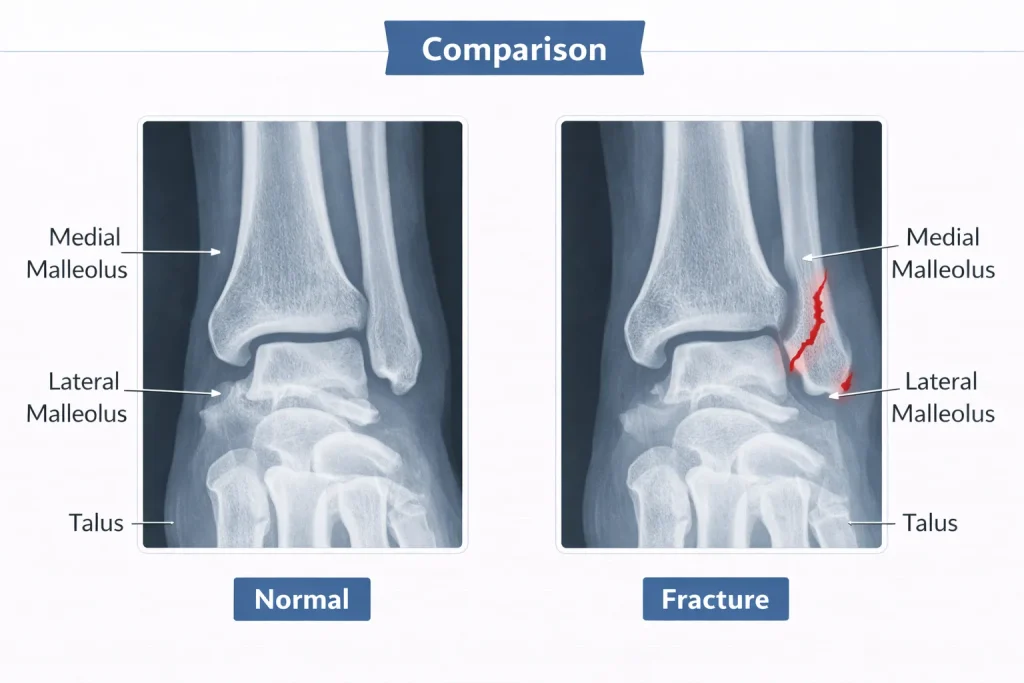
An ankle fracture refers to a break in one or more bones forming the ankle joint. The joint is composed of:
- The tibia (main shin bone)
- The fibula (outer lower leg bone)
- The talus (bone connecting leg to foot)
The bony prominences felt on either side of the ankle are called malleoli:
- Medial malleolus (inner side)
- Lateral malleolus (outer side)
- Posterior malleolus (back part of tibia)
An ankle fracture may involve one, two, or all three malleoli. The severity depends not only on the bone break but also on ligament damage and joint stability.
It is important to differentiate between an ankle sprain and a fracture. While sprains involve ligament injury, fractures involve bone disruption and require imaging confirmation.
Anatomy of the Ankle Joint
Understanding ankle anatomy helps explain why certain fractures require surgery.
The ankle joint (talocrural joint) is a hinge joint that allows:
- Dorsiflexion (lifting foot upward)
- Plantarflexion (pointing foot downward)
The stability of the ankle depends on:
- The deltoid ligament on the medial side
- Lateral ligament complex (ATFL, CFL, PTFL)
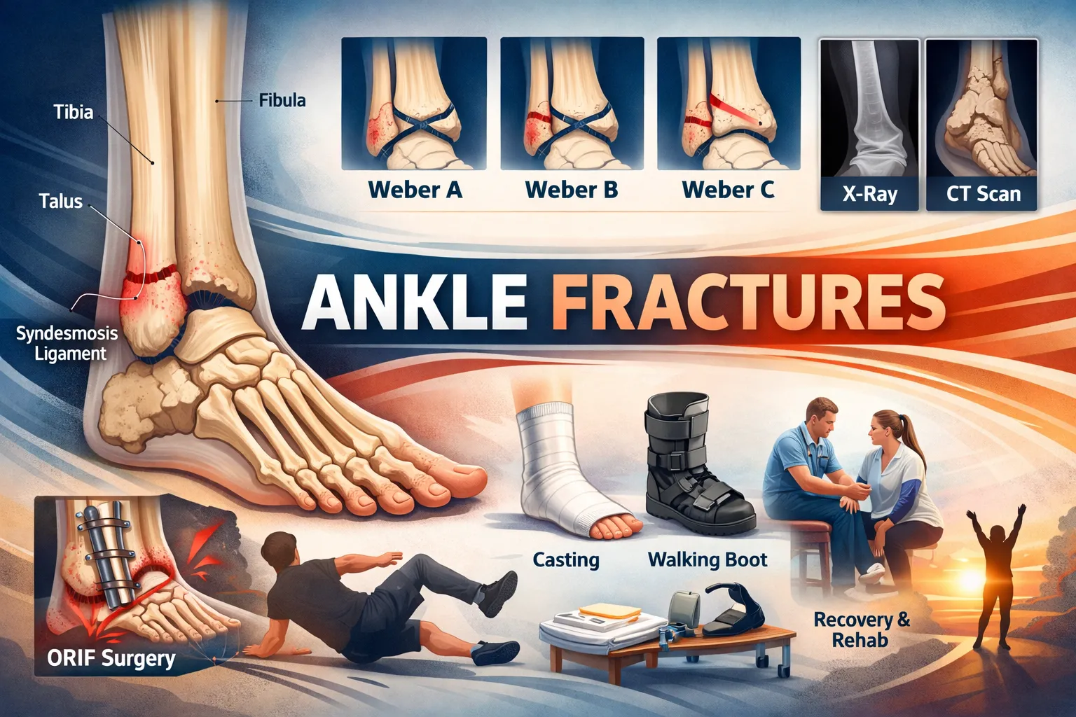
- Syndesmotic ligaments connecting the tibia and fibula
Even a small misalignment in the ankle joint can lead to abnormal load distribution and long-term arthritis. That is why anatomical reduction is critical in unstable fractures.
Types of Ankle Fractures
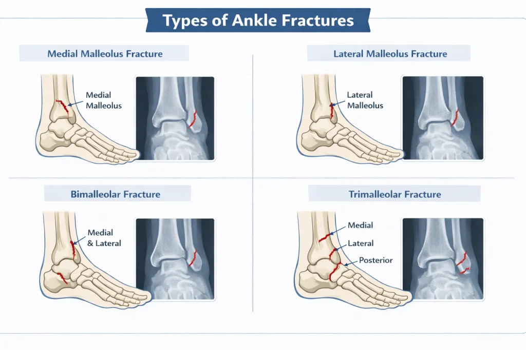
Ankle fractures are categorized based on which part of the ankle is broken. The fracture pattern determines stability and treatment.
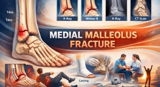
Medial Malleolus Fracture
Medial Malleolus Fracture occurs on the inner side of the ankle and may result from twisting injuries or high-impact trauma.
Common features include:
- Inner ankle pain
- Tenderness along the tibia
- Possible vertical or transverse fracture pattern
Stable fractures may heal with casting, while displaced fractures often require surgical fixation.
Lateral Malleolus Fracture (Distal Fibula Fracture)
Lateral Malleolus Fracture is the most common ankle fracture globally. It involves the lower part of the fibula.
Key considerations:
- May be stable or unstable
- Stability depends on syndesmotic involvement
- Often associated with ligament injury
Stable fractures can be treated conservatively, but unstable ones require ORIF.
Bimalleolar Fracture
A bimalleolar fracture involves both medial and lateral malleoli. Because two stabilizing structures are broken, the ankle joint is usually unstable.
Typical management includes:
- Surgical realignment
- Plate and screw fixation
- Structured rehabilitation
Trimalleolar Fracture
This injury affects:
- Medial malleolus
- Lateral malleolus
- Posterior malleolus
It often occurs after high-energy trauma and frequently requires surgical stabilization to restore joint alignment.
Ankle Fracture Classification Systems
Classification systems help doctors predict instability and guide treatment.
Weber Classification
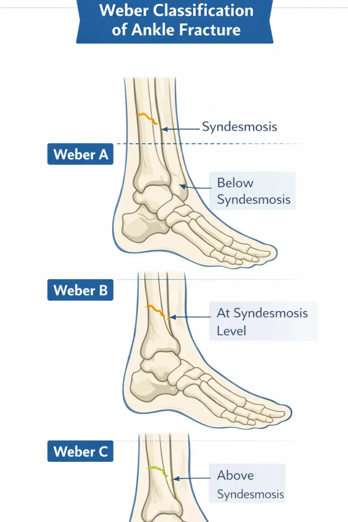
The Weber classification is based on the level of fibula fracture relative to the syndesmosis:
- Type A – Below the syndesmosis (usually stable)
- Type B – At the syndesmosis level (variable stability)
- Type C – Above syndesmosis (usually unstable)
Weber C injuries often involve ligament disruption and need surgical fixation.
Lauge-Hansen Classification
This classification describes the mechanism of injury based on foot position and force direction.
Common patterns include:
- Supination-External Rotation (most common globally)
- Pronation-External Rotation
- Supination-Adduction
While more academic in nature, it helps predict associated ligament damage.
Causes of Ankle Fractures
Ankle fractures occur due to:
- Twisting injury during walking or running
- Sports trauma
- Road traffic accidents
- Fall from height
- Osteoporotic fragility fractures
In India and globally, sports injuries and urban accidents are leading causes.
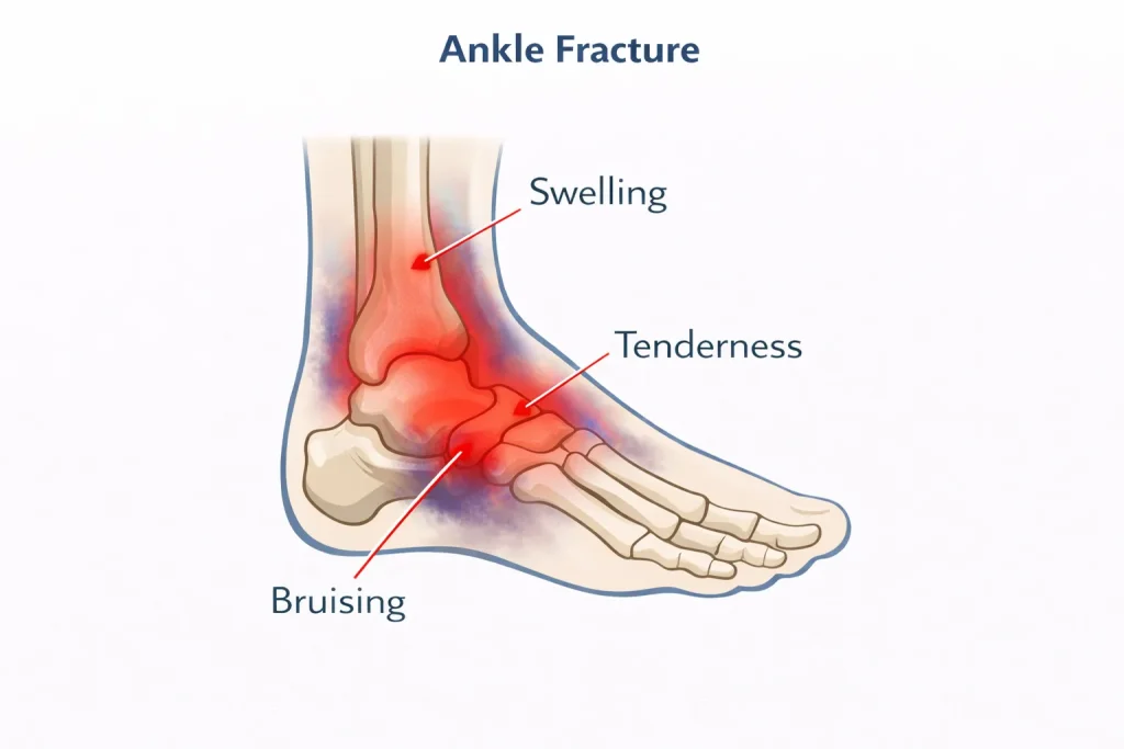
Symptoms of Ankle Fracture
Symptoms typically appear immediately after injury and vary based on severity.
Common symptoms include:
- Sudden ankle pain
- Swelling around the joint
- Bruising or discoloration
- Difficulty bearing weight
- Deformity in severe cases
Persistent pain after a twisting injury should not be ignored, even if swelling seems mild.
Diagnosis of Ankle Fracture
Accurate diagnosis is essential to determine stability.
Clinical Examination
Doctors assess:
- Bone tenderness
- Swelling
- Deformity
- Neurovascular status
Imaging
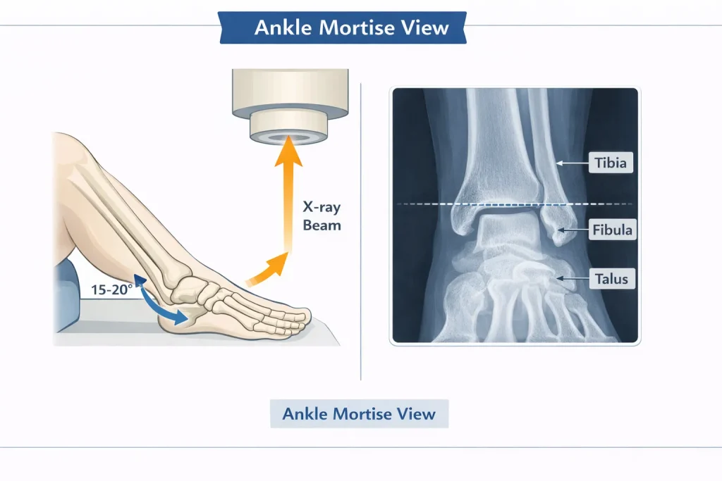
X-ray
Standard views include:
- AP view
- Lateral view
- Mortise view
The mortise view is critical to evaluate joint alignment.
CT Scan
A CT scan is recommended when:
- Posterior malleolus is involved
- Fracture pattern is complex
- Surgical planning is required
Treatment of Ankle Fractures
Treatment depends on stability, displacement, patient age, and activity demands.
Non-Surgical Treatment
Indicated for stable fractures.
Management includes:
- Short leg cast or walking boot
- Non-weight bearing for 4 – 6 weeks
- Gradual rehabilitation
Surgical Treatment (ORIF)
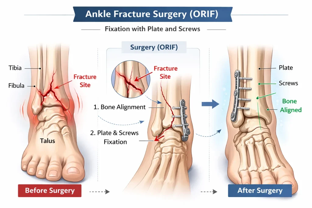
ORIF (Open Reduction and Internal Fixation) is required when:
- Fractures are displaced
- The joint is unstable
- Syndesmosis is disrupted
- Bimalleolar or trimalleolar fractures occur
Surgery restores alignment using:
- Plates
- Screws
- Syndesmotic fixation
Precise anatomical reduction reduces arthritis risk.
Recovery Timeline After Ankle Fracture
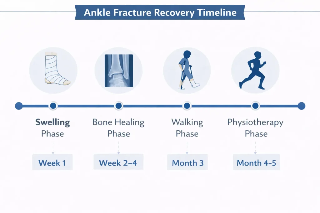
Recovery varies depending on fracture type.
| Fracture Type | Weight Bearing | Full Recovery |
|---|---|---|
| Stable fracture | 4 – 6 weeks | 8 – 10 weeks |
| Bimalleolar | 6 – 8 weeks | 3 – 4 months |
| Trimalleolar | 8 – 10 weeks | 4 – 6 months |
| Complex fracture | 10 – 12 weeks | 6+ months |
Rehabilitation includes:
- Range of motion exercises
- Strength training
- Balance correction
- Gait training
Swelling may persist for months even after bone healing.
Complications of Ankle Fractures
Complications may occur if alignment is not restored properly.
Possible complications include:
- Post-traumatic arthritis
- Malunion
- Nonunion
- Infection
- Chronic stiffness
Early expert intervention significantly reduces these risks.
Consultation & Global Care with Dr Divya Ahuja
If you have sustained an ankle injury and need expert evaluation, early consultation can prevent long-term complications.
Dr Divya Ahuja provides advanced orthopedic care for:
- Complex ankle fractures
- Bimalleolar and trimalleolar injuries
- Fracture-dislocations
- Revision surgery cases
Why choose India for orthopedic treatment?
- International standard surgical techniques
- Significantly lower treatment cost
- Advanced imaging and rehabilitation facilities
- Personalized patient care
International patients can access:
- Online consultation
- Treatment planning
- Surgical scheduling
- Post-operative rehabilitation guidance
Early expert intervention ensures better joint stability, faster recovery, and lower arthritis risk.
FAQs
How long does a medial malleolus fracture take to heal?
A medial malleolus fracture typically heals within 6 – 8 weeks if stable and treated with casting. Surgical cases may take 3 – 4 months for full recovery, including rehabilitation. Healing time varies based on age, bone health, and fracture displacement.
What is the recovery time for a bimalleolar ankle fracture?
Bimalleolar ankle fracture recovery usually takes 3 – 4 months. Patients remain non-weight-bearing for about 6 – 8 weeks, followed by progressive physiotherapy. Full return to sports or heavy work may take up to 6 months.
Can a lateral malleolus fracture heal without surgery?
A lateral malleolus fracture can heal without surgery if it is stable and not displaced. However, unstable fractures or those involving syndesmotic injury often require surgical fixation to prevent long-term ankle instability and arthritis.
How long does ankle fracture swelling last after surgery?
Swelling after ankle fracture surgery can last for several weeks. Mild swelling may persist for 3 – 6 months. Elevation, compression, and physiotherapy help reduce swelling and improve circulation during recovery.
What is Weber B fracture treatment?
Weber B fracture treatment depends on stability. Stable Weber B fractures may be treated with casting, while unstable cases require ORIF surgery. Proper evaluation with stress X-rays determines the need for surgery.
When can I walk after ankle fracture surgery?
Most patients begin partial weight-bearing 6 weeks after ankle fracture surgery, depending on healing. Full weight-bearing typically starts after radiographic confirmation of bone union.
Does an ankle fracture always need a cast?
Not all ankle fractures require a traditional cast. Some stable fractures may be managed using a removable walking boot. However, immobilization is essential for proper healing.